Neoplastic diseases
Hepatic epithelial cell origin tumors
Hepatoma/hepatocellular adenoma/hepatocellular carcinoma
Although the diagnosis of primary hepatic neoplasia can be cytologically challenging, the use of a complex logistic regression model has shown that cytologic findings can be used to diagnose hepatocellular carcinoma (Stockhaus et al., 2004).
This study found that hepatocytes with a high N:C ratio, larger cell diameters, increased numbers of nucleoli per nucleus, small numbers of cytoplasmic vacuoles, and small numbers of lymphocytes intermixed with hepatocytes were associated with hepatocellular carcinoma. Additionally, primary hepatic tumors will commonly display a more uniform population of abnormal hepatocytes in contrast to the mixture of dysplastic hepatocytes and cytologically unremarkable cells seen with other pathologic processes. Another canine study found that dissociated hepatocytes, acinar or palisading arrangements of neoplastic cells, and the presence of naked nuclei and capillaries, when seen with mild anisocytosis, anisokaryosis, multinucleation, and increased N:C ratio, were useful for diagnosing well-differentiated hepatocellular carcinoma (Figures 9.27, 9.34–9.36, Masserdotti Drigo, 2012).Hepatocellular adenomas and carcinomas have been described in both dogs and cats (Trigo et al., 1982; Balkman, 2009). In the dog, hepatocellular carcinoma is the most common primary liver tumor. The metastatic rate has been reported to be as high as 60% with common sites of metastasis being the lung, local lymph node, and peritoneum. Even with common metastasis, treatment has been associated with survival times of approximately 4 years (Balkman, 2009). In the cat, hepatocellular adenoma is more common.
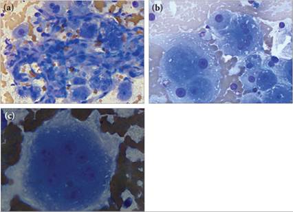
Figures 9.34a–c Liver aspirate from a Labrador Retriever with multiple hepatic masses.
(a) This slide contains a few more normal looking hepatocytes, often enmeshed in a network of capillaries. (b) Cytologically, there was a continuum of cell morphology from large cohesively clustered cells, which have pink granular to basophilic cytoplasm, to those with more basophilic cytoplasm and poorly distinct cell borders (giving a naked nuclei appearance). Marked nuclear pleomorphism was also noted. (c) Rarely, very large multinucleate cells with marked nuclear and nucleolar variability are also seen. These features have been associated with hepatocellular carcinoma. In conjunction with a history of a hepatic mass noted on ultrasound, the slides are suggestive of hepatocellular carcinoma (a and b Wright–Giemsa, 500? magnification; c, 1,000? magnification).
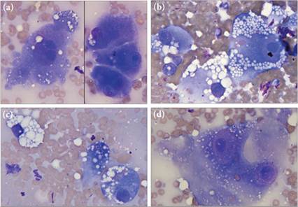
Figures 9.35a–d Liver aspirate from a Cocker Spaniel with multiple hepatic masses. Cytologically, many features suggesting hepatocellular carcinoma are present. (a) Anisocytosis, anisokaryosis, binucleation, and multiple nucleoli are present. (b) The neoplastic cell in the upper right corner has a nucleolus approximately the size of the nuclei in the other two intact hepatocytes. Most of the hepatocytes also contain abundant clear cytoplasmic vacuolation, consistent with lipid. The fuchsia material noted in the image is consistent with ultrasound gel. (c) Note the highly variable amount of clear distinct vacuolation. Without a progression of cells from minimally vacuolated to highly vacuolated, the cell in the upper left corner would be challenging to recognize as a hepatocyte. (d) Markedly cytomegalic cells are present. Again, nuclei are large and contain impressively large dark nucleoli (Wright–Giemsa, 1,000? magnification).
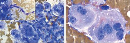
Figures 9.36a,b This dog had multiple liver nodules on ultrasound and several features suggestive of hepatocellular carcinoma on liver cytology. (a) Capillaries are commonly seen within hepatocyte clusters. (b) Many multinucleated cells with three to seven nuclei are present. Cell borders of many hepatocytes are also poorly distinct, suggesting a fragile population of cells. Note the multiple and variable size of nucleoli (Wright–Giemsa, 1,000? magnification).