Organization of the nervous system
For the purpose of presentation, the nervous system is generally divided into two major divisions: the central nervous system (CNS) and the peripheral nervous system (PNS) (Fig. 8.2).
The CNS includes the brain and spinal cord. These are located in the dorsal body cavity and are encased in the skull and vertebrae. The CNS includes not only neurons but also blood vessels, connective tissue, and supportive cells. The CNS is responsible for the integrative function in which sensory information from both inside and outside the body is processed and the appropriate response is generated.The PNS includes all the neurons outside the CNS. These include the spinal nerves that carry impulses to and from the spinal cord and the cranial nerves that carry impulses to and from the brain. Peripheral nerves consist of the neurons and associated blood vessels and connective tissue that lie outside of the CNS.
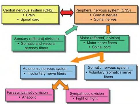
Fig. 8.2. Organization of the nervous system. The two main divisions of the nervous system include the central and peripheral nervous system. The peripheral nervous system has a sensory division that carries signals toward, and a motor division that carries signals away from, the central nervous system, respectively. The motor division is further divided into the autonomic component that controls involuntary functions, such as gastrointestinal tract motility and heart rate, and a somatic component that controls skeletal muscle. The autonomic nervous system is made of the parasympathetic and sympathetic divisions.
The PNS is further divided into the sensory, or afferent (ad = to + ferre = to carry), and motor, or efferent (ex = from), division. The sensory division consists of sensory neurons located throughout the body that project to the brain and spinal cord.
This sensory input is carried in the sensory division of the PNS. Sensory input originates from receptors, which are specialized structures that detect changes in either the internal or external environment. These receptors can be as simple as the dendrite of a neuron, or they can consist of an organ adapted to detect a specialized type of information such as the Golgi tendon organ, which detects the stretch of the tendons. The motor response generated via the integrative function of the CNS is carried to either a muscle or endocrine gland by the motor division of the PNS.The motor division of the PNS is further divided into two parts: somatic nervous system and autonomic nervous system. The somatic nervous system controls skeletal muscle contractions and is under voluntary control. Therefore, an animal can consciously control the somatic nervous system.
The autonomic nervous system, also called the visceral motor system, controls smooth muscle, cardiac muscle, and glandular secretions. In contrast to the somatic nervous system, the autonomic nervous system is under involuntary control, meaning that its regulation generally occurs at the subconscious level. An animal does not have to consciously control the dilation or constriction of a blood vessel in the skin in response to heat. Instead, this happens automatically— hence the name autonomic nervous system. The autonomic nervous system includes the sympathetic and parasympathetic divisions. These two divisions generally have an antagonistic effect on various functions. For example, stimulation of the sympathetic nervous system will increase heart rate, whereas stimulation of the parasympathetic nervous system will decrease heart rate.
The neuron
The nervous system consists of neurons and supportive cells. Neurons are excitable cells that are able to transmit an electrical impulse along their length. Supportive cells are responsible for making the myelin sheath surrounding many neurons, providing nutrients, as well as performing a phagocytic role.
Neurons are highly specialized cells that can respond to stimuli, and produce an impulse, and transmit that information to a distant site (Fig. 8.3). Neurons come in many sizes and shapes. They have a long life span and are generally considered amitotic, meaning that they no longer divide. However, recent evidence has revealed that within certain sites in the brain, neurons do reproduce. It has been demonstrated in songbirds that the number of neurons in regions of the brain associated with the production of songs increases in the spring as the birds increase their repertoire in preparation for mating. Similarly, the number of neurons in sites within the human brain, such as
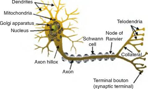
Fig. 8.3. A typical neuron. A typical neuron has a cell body (soma) that contains various organelles. The dendrites act as afferent fibers carrying signals to the soma. A neuron typically has a single axon that is the efferent fiber carrying signals away from the cell body. Many times, the axon is surrounded by a myelin sheath that acts to insulate the process. In the periphery, Schwann cells make the myelin, whereas in the central nervous system, it is made by oligodendrocytes. The space between adjacent Schwann cells is called the node of Ranvier.
the hippocampus, has been shown to increase under certain conditions. It is assumed this happens in other species.
Cell body
The cell body, or soma, consists of a large, round nucleus 5-10 μm in diameter surrounded by cytoplasm, also called perikaryon (karyon = nucleus). Within the cytoplasm are the normal cell organelles except for the lack of centrioles that are responsible for formation of the mitotic spindle associated with cell division.
The cytoplasm contains free ribosomes, smooth and rough endoplasmic reticula (ERs), mitochondria, and Golgi apparatus. The rough ER is also known as Nissl substance that stains darkly in the presence of basic dyes called Nissl stains.
The rough ER is the major site of protein synthesis destined for insertion into the membrane of the cell or an organelle. Free ribosomes are responsible for the synthesis of proteins destined for the cytosol. Smooth ER can be continuous with rough ER and acts as a site where newly synthesized proteins are folded into their three-dimensional structure. Smooth ER can also regulate cytosolic concentrations of ions such as calcium.Also within the cytosol is an internal scaffolding called the cytoskeleton. The cytoskeleton consists of several components. The largest are the microtubules measuring about 20 nm in diameter and running longitudinally down the neurites (axons and dendrites). They are formed through polymerization of molecules of the protein tubulin. Associated with the microtubules is another class of proteins called microtubule- associated proteins (MAPs). These proteins help anchor microtubles to other parts of the neuron and to each other. The second component of the cytoskeleton is neurofilaments, which are called intermediate fibers in other cell types, measuring IOnm in diameter. They consist of multiple subunits connected end to end. Each subunit consists of three proteins interwoven together. A third component is the microfilaments measuring 5nm in diameter. They consist of two braided strands, each of which is made of polymers of the protein actin. Microfilaments not only run longitudinally down the neurites but are also anchored to the inside of the cell membrane.
Dendrites
The term dendrite is derived from the Greek word for "tree," and as such, they look like the branches of a tree originating from the cell body. All of the dendrites collectively make up the dendritic tree. The dendrites on some neurons also have on their surface dendritic spines. Dendrites act as the receptive region of the neuron. The combination of the dendritic tree and dendritic spines makes for a large surface area that facilitates this function. The cytoplasm of the dendrites resembles that of the soma.
As discussed further, neurons can be classified based on dendrites.Axon
While the structures discussed earlier are common to most cells, the axon is unique to neurons. The axon is specialized to allow an impulse to be transmitted along its length and thus carried from one location to another. A long axon is sometimes called a nerve fiber. A bundle of axons within the CNS is called a tract; it is called a nerve in the periphery.
A neuron has a single axon that originates from the soma in a region called the axon hillox. This is where the nerve impulse originates and, therefore, is sometimes called the trigger zone. The axon is unique in that it contains no rough ER; it contains few, if any, free ribosomes; and its membrane has a different protein composition from that of the soma.
While the axoplasm (cytoplasm inside the axon) contains neurofibrils, neurotubules, lysosomes, mitochondria, and small vesicles, it lacks rough ER and ribosomes. Therefore, protein synthesis does not occur in the axon. Instead, proteins must be synthesized in the soma and transported along the axon.
Axons may extend less than a millimeter or over a meter in length. Axons typically branch, forming collaterals that enable one neuron to communicate with several other sites. Occasionally, an axon collateral may communicate with the same neuron from which it originated, thus forming a recurrent collateral. The diameter of an axon can range from less than 1 μm to as large as 1 mm. The thicker the axon, the faster the speed of conduction of the nerve impulse down its length.
The nerve fibers of many nerves are covered with a whitish, fatty sheath called myelin. Myelin acts to protect and insulate the axon and increases the speed of conduction of the impulse. Whereas the speed of conduction may be 1 m/s in unmyelinated fibers, it can be 150 m/s in myelinated fibers. The dendrites are always unmyelinated.
In the PNS, the myelin is produced by the Schwann cells. The Schwann cell spirals around the axon, producing many concentric circles enclosing the axon and forming the myelin sheath.
During the spiraling process, the nucleus and cytoplasm of the Schwann cell gets squeezed to the outer layer of the cell and appears as a bulge on the outer surface. The outer layer is the neurilemma. Occasionally, the Schwann cell does not spiral around the axon, but instead encloses many axons at one time in what look like indentations on its surface. Such axons are said to be unmyelinated. Adjacent Schwann cells do not touch one another, but instead form a space called the node of Ranvier, or neurofibral nodes, in which the axonal membrane is exposed.In the CNS, myelin is produced by another type of cell called an oligodendrocyte. Oligodendrocytes are a type of glial, or supportive, cell in the CNS. Instead of spiraling around the axon, the oligodendrocytes form end feet that surround the axon and form the myelin sheath. One oligodendrocyte can thereby myelinate many axons, whereas a Schwann cell myelinates only a single axon. In the CNS, areas containing myelinated fibers are referred to as white matter and generally consist of fiber tracts. Areas containing cell bodies are referred to as gray matter; collections of cell bodies are called nuclei.
The collaterals off the main axon trunk end in a series of fine extensions called telodendria. A collection of telodendria is called a terminal arbor. The telodendria end in a knoblike structure called the axon terminal, terminal bouton, or synaptic knob. Microtubules do not extend into the terminal, but the terminal will typically contain synaptic vesicles. Synaptic vesicles are small membrane-bound spheres measuring 50 nm in diameter and containing quanta of neurotransmitters. The axon terminal will end at another neuron or cell such as an endocrine gland or muscle cell.
Synapse
Where the axon terminal meets another cell is called the synapse. It consists of a presynaptic membrane, a synaptic cleft, and a postsynaptic membrane. The axon terminal will typically contain synaptic vesicles. The average neuron forms approximately 1000 synaptic junctions.
The synaptic terminal of one neuron can synapse on any part of an adjacent neuron. Therefore, this can create synapses such as axoaxonic, axodendritic, and axosomatic. When a neuron synapses on a skeletal muscle cell, it creates a specialized junction called a neuromuscular junction. If the neuron synapses with a gland, it creates a neuroglandular synapse.
There are two types of synapses, electrical and chemical, which have different functional properties (Table 8.1). While chemical synapses are much more common between neurons in the mammalian and avian brain, electrical synapses are common between nonneural cells such as glial cells, epithelial cells, smooth and cardiac muscle cells, liver cells, and some glandular cells.
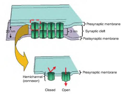
Fig. 8.4. An electrical synapse. An electrical synapse consists of a gap junction between two adjacent cells. The space between the presynaptic and postsynaptic cells contains channels called connexons, each composed of six protein subunits called connexins. The connexon forms a cytoplasmic connection between adjacent cells allowing ions and small molecules to pass between both cells. The connexins can tilt toward each other, closing the channel.
| Table 8.1. Electrical versus chemical synapses. | |||||
| Type of Synapse | Distance within Synaptic Cleft | Components of Synapse | Agent of Transmission | Synaptic Delay | Direction of Transmission |
| Chemical | 20-40 nm | Synaptic vesicles, active zone, postsynaptic receptors | Chemical | 1-5 ms | Unidirectional |
| Electrical | 3.5 nm | Gap-junction channels | Electrical | Virtually absent | Bidirectional |
In electrical synapses, the pre- and postsynaptic cells communicate through special channels called gap junctions (Fig. 8.4). These junctions provide a channel between the cytoplasm of the adjacent cells. Gap junctions consist of a pair of hemichannels, with one associated with the presynaptic membrane and the other with the postsynaptic membrane. Each hemichannel consists of specialized proteins called connexins. Six connexins combine to form a channel called a connexon through which ions can pass from the cytoplasm of one cell to the cytoplasm of another cell. The pore formed within the connexon is about 2 nm in diameter, making it one of the largest known, and is of sufficient size to allow small organic molecules to pass. It appears that the connexins can tilt toward one another to close the channel.
The electrical synapse allows an action potential to pass from one cell to another with virtually no delay. This is beneficial when it is necessary for a signal to pass rapidly to another cell, and for that signal to not be modified.
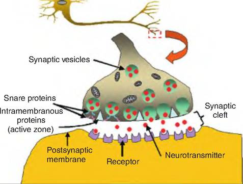
Fig. 8.5. A chemical synapse. A chemical synapse consists of synaptic membrane, synaptic cleft, and a postsynaptic membrane. Since the nerve impulse cannot jump across the synaptic cleft, a neurotransmitter is released, which carries the signal to the postsynaptic cell. The active zone of the presynaptic cell contains intramembranous proteins, thought to be calcium channels. When the neuron is depolarized, calcium enters through the calcium channels and causes the synaptic vesicles to bind to the presynaptic membrane and release their contents through a process of exocytosis. SNARE (soluble N-ethylmaleimide-sensitive proteins) attaches synaptic vesicles to the presynaptic membrane.
The chemical synapse is characterized by a synaptic cleft 20-50nm wide (Fig. 8.5). Within the synaptic cleft is a fibrous protein matrix to help the two cells adhere to one another. An electrical signal cannot cross the synaptic cleft, so a chemical substance called a neurotransmitter carries the signal to the postsynaptic cell. The neurotransmitter is stored in the synaptic vesicles. The concentration of neurotransmitter can be 10,000 times higher than in the cytosol. This is accomplished by a countertransport system in which a molecule of transmitter enters the vesicle in exchange for a H+ ion (Fig. 8.6).
Also found on the presynaptic membrane are pyramid-shaped proteins projecting into the cytoplasm. These proteins, and the associated cell membrane, make the active zone. The active zone is the site of neurotransmitter release. The pyramid-shaped
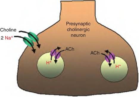
Fig. 8.6. Pumping neurotransmitters. Neurotransmitters are actively concentrated into the synaptic vesicles, using a countertransport system. The vesicle membrane has an H+-ATP pump that loads the vesicle with H+. The neurotransmitter then enters the vesicle in exchange for a molecule of H+. In addition, there is a transport system on the presynaptic neuron membrane that transports either the neurotransmitter, or its precursor, into the cell. In this example, choline, the precursor Ofacetylcholine (ACh), is actively cotransported into the cell with Na+.
proteins associated with the active zone are believed to be calcium channels.
Classification of neurons
Neurons can be classified based on several characteristics. These include the structural classifications based on the number of neurites that extend from the soma or the length of the axon, the function, or the neurotransmitter they contain.
Classification based on neurite number
Based on neurite number, neurons can be classified as unipolar, bipolar, pseudounipolar, or multipolar (Fig. 8.7). Unipolar neurons are the simplest nerve cells, and they have a single process. They are found in the autonomic nervous system. Bipolar neurons have an oval-shaped soma from which two processes emerge: the dendrite and the axon. Many sensory neurons are bipolar, such as those in the retina of the eye and olfactory epithelium of the nose. Pseudounipolar cells serve as mechanoreceptors that sense touch, pressure, and pain. The pseudounipolar neuron develops embryologically as a bipolar neuron with two processes, but eventually, these two processes fuse into a single axon that emerges from the soma. The axon then divides into two segments, with one going to the periphery and one going to the spinal cord. The pre-
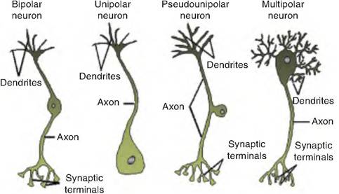
Fig. 8.7. Classification of neurons. Anaxonic neurons lack an axon. Bipolar neurons have one dendrite and one axon originating from the cell body. In unipolar neurons, the dendrite and axon merge into a single process having a dendritic and axonic component. Pseudounipolar neurons have a small process coming off at the cell body and leading to the axon. In multipolar neurons, there are many dendrites but a single axon originating from the cell body.
dominate type of neuron is the multipolar neuron, which has a single axon and generally has many dendrites.
Classification based on axon length
Classifications based on axon length include Golgi type I and Golgi type II neurons. Golgi type I neurons have long axons and are considered projection neurons since they carry signals to other sites. An example would be pyramidal cells with the cell bodies located in the cerebral cortex and whose axons extend to the spinal cord. Golgi type II neurons have short axons and are involved in local circuits. Examples include stellate or basket neurons in the cerebellum.
Classification based on function
Based on function, neurons are classified the following ways: (1) sensory or afferent neurons, (2) motor or efferent neurons, and (3) interneurons or association neurons. Sensory neurons respond to sensory stimuli and transmit that information to the nervous system, with most information being carried to the CNS. Most sensory neurons are pseudounipolar, and their cell bodies are located in the dorsal root ganglion of the spinal nerves. Motor neurons transmit signals from the brain or spinal cord to the muscles or glands. Their cell bodies are located in the CNS. Interneurons are the largest class of neurons—constituting 99% of all neurons, including all those neurons that are not sensory or motor—and are generally multipolar. This class can be divided into two groups. Relay or projection interneurons have long axons and transmit
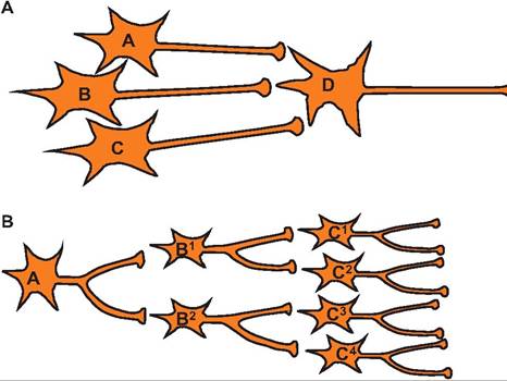
Fig. 8.8. Diverging and converging neuronal pools. Interneurons are involved in making neuronal pools. (A) Convergence occurs when several interneurons synapse on a single neuron.
(B) Divergence occurs when a single interneuron synapses on more than one interneuron.
signals over considerable distances, such as the dorsal columns located in the spinal cord. Local interneurons have short axons and are involved in processing information in localized circuits, such as the horizontal cells in the retina or stellate cells that inhibit Purkinje cells in the cerebellum.
Interneurons, sometimes called association neurons, are the most numerous of all neuronal types. Mostly located in the brain and spinal cord, some are found in autonomic ganglia. They function to distribute sensory information and coordinate motor activity. Interneurons produce patterns of connections such as divergence and convergence (Fig. 8.8). Information coming from a single source and spreading to multiple neurons is known as divergence, whereas input from multiple neurons synapsing on a single interneuron is called convergence.
Classification based on neurotransmitter
Finally, neurons can be classified according to the neurotransmitter they release. In the case of motor neurons, which innervate skeletal muscle, they all release acetylcholine (ACh) and are called cholinergic neurons. Neurons that release serotonin, such as 5-hydroxytryptamine (5-HT), are called serotonergic neurons. An example would be those neurons in the raphe nucleus of the brain stem.