Plasma cell tumors
Plasma cell tumors in the spleen include multiple myelomas (mainly in cats) and extramedullary plasmacytomas. Diffuse enlargement of spleen and a massive presence of plasma cells at different stages of differentiation are the main features.
Cells may exhibit characteristics of malignancy and binucleated cells are frequent (Figure 5.72). Monoclonal increases in globulin concentration may be detected by serum protein electrophoresis. 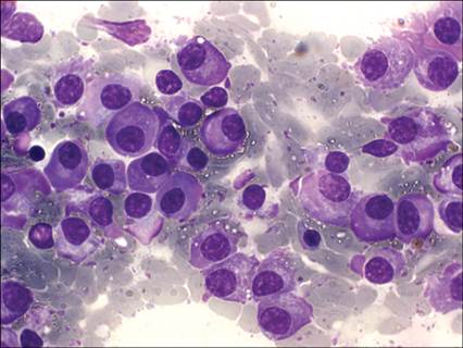
Figure 5.72 Dog, spleen, FNA. Plasma cell tumor. A prevalent population of medium-sized round cells with abundant cytoplasm, often irregularly eosinophilic, eccentric round nuclei with coarse to clumped chromatin and no visible nucleoli (May–Grünwald–Giemsa, 1,000? magnification).
Histiocytic disorders
The spleen is a common site of malignant histiocytic tumors. Two different types of histiocytic sarcomas may be distinguished based on cytomorphology and immunoreactivity. Disseminated histiocytic sarcoma derives from a clonal expansion of splenic dendritic cells with a CD1+, CD11c+, CD11d−, MHC-II+ immunophenotype (Affolter & Moore, 2002). Neoplastic cells are large and discrete, often with abundant, clear cytoplasm, indistinct cell borders, and cytoplasmic vacuoles. The nucleus is oval to convoluted with finely reticulated or irregularly clumped chromatin (Figures 5.73). Binucleated and multinucleated giant cells are frequent and characteristics of malignancy are prominent including severe anisocytosis and anisokaryosis. Hemophagocytic histiocytic sarcoma derives from macrophages that are immunopositive for CD11d, while expression of CD1 and MHC-II is low and CD11c is generally negative (Moore et al., 2006). This neoplasm has a poor prognosis.
A mild to moderate anemia is often present. Neoplastic cells are large with foamy cytoplasm and contain phagocytized erythrocytes and/or hemosiderin (Figures 5.74–5.76). The detection of erythrophagocytosis in neoplastic cells supports the diagnosis of hemophagocytic histiocytic sarcoma; however, several other round cell tumors (mastocytoma, lymphoma, plasma cell tumors, osteosarcoma, megakaryocytic leukemia; Figures 5.77–5.79) and some sarcomas (e.g., hemangiosarcoma, osteosarcoma) have been demonstrated to occasionally exhibit erythrophagocytosis (Barger et al., 2012); thus this feature must be interpreted with caution. 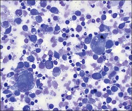
Figure 5.73 Dog, spleen, FNA. Disseminated histiocytic sarcoma. Large histiocytoid cells, with vacuolated cytoplasm intermingled with lymphoid cells and neutrophils (Wright–Giemsa, 500? magnification).
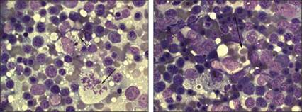
Figure 5.74 Large histiocytoid neoplastic cells and a huge atypical mitotic figure are found in a background of severe extramedullary hematopoiesis and erythrophagocytosis (arrows).Figure 5.75 Intense recent erythrophagocytosis (arrows) in a neoplastic macrophage. Some histiocytoid cells are also seen (May–Grünwald–Giemsa, 1,000? magnification).
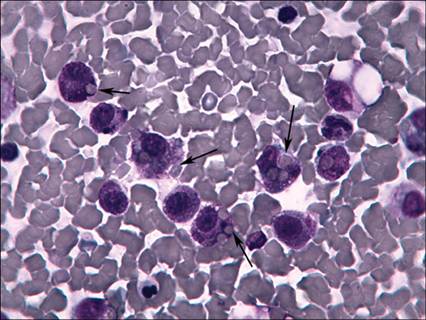
Figure 5.76 Cat, spleen, FNA. Mast cell tumor with erythrophagocytosis (arrows) (May–Grünwald–Giemsa, 1,000? magnification) (courtesy M. Gelain).
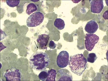
Figure 5.77 Dog, spleen, FNA. Diffuse large B-cell lymphoma. Recent erythrophagocytosis (arrows) in a neoplastic blast cell (May–Grünwald–Giemsa, 2,000? magnification).
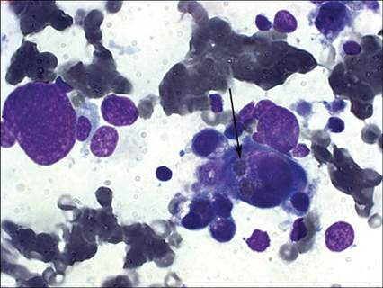
Figure 5.78 Dog, spleen, FNA.
Acute megakaryoblastic leukemia. Recent erythrophagocytosis (arrow) in a neoplastic cell (May–Grünwald–Giemsa, 1,000? magnification).
Mast cell tumors
Although mast cells are a normal resident cell population in spleen, their percentage may increase in reactive conditions and with metastatic mast cell tumors. The detection of a high number of mast cells in the spleen of either dogs or cats with mast cell tumors is associated with a worse prognosis (Book et al., 2011). Splenic infiltration from cutaneous mastocytoma is generally associated with splenomegaly and has been reported in 37% of a cohort of 19 dogs (Book et al., 2011) and even more commonly in feline mast cell tumors (Spangler & Culbertson, 1992). Determining if the number of mast cells in a splenic aspirate is increased can be difficult. Stefanello et al. (2009) suggested the following aspects to define splenic infiltration: (1) presence of aggregates of well-differentiated mast cells; (2) presence of a large number of well-differentiated mast cells (Figure 5.79); or (3) presence of mast cells with atypical morphology (poor granulation or pleomorphism). Poorly granulated mast cells may be frequently found in cats, mainly when visceral mastocytosis is present. In these cases, care should be taken to differentiate neoplastic mast cells from other round cell tumors.
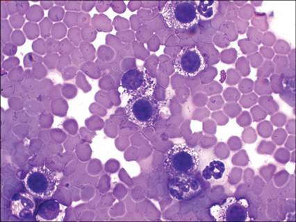
Figure 5.79 Dog, spleen, FNA. Well-differentiated mast cell tumor. Presence of an abundant population of well-differentiated mast cells (Diff-Quik®, 1,000? magnification) (courtesy D.M. Lupi).