Lymphoma
The distinction between primary splenic lymphoma and metastatic lymphoma is not always possible, but a high percentage of primary splenic lymphomas are low-grade B-cell lymphomas often arising from the marginal or the mantle zone, and systemic involvement is limited.
Detection of an increased percentage of blasts should be interpreted with care, since blasts are increased in hyperplastic and reactive conditions. Detection of more than 40% blasts in a splenic FNB is suggestive of lymphoid neoplasia. If necessary, PARR may help reach a more definitive diagnosis (Williams et al., 2006). Other papers suggest considering a splenic involvement when there are >5% blasts in dogs with a previous diagnosis of multicentric lymphoma (Nerschbach et al., 2016).Histology is necessary to further subtype lymphomas of the spleen and may provide some prognostic information. Marginal zone lymphomas are nodular (Figures 5.69, 5.70) or diffuse and have a more favorable prognosis in dogs than marginal zone lymphoma in the lymph node (Stefanello et al., 2011). Mantle cell lymphomas have been reported in spleen from both dogs and cats and have a good prognosis (Valli, 2007). Hepatosplenic lymphoma is a rare and aggressive T cell lymphoma variant described cytologically as medium-/large-sized cells that often have fine magenta cytoplasmic granules and variably shaped nuclei. High-grade, diffuse large B-cell lymphoma commonly infiltrates the spleen and progresses from multifocal to coalescing lesions, with variable response to therapy (Valli, 2007). In dogs, T-cell chronic lymphocytic leukemia (CLL) often arises from the spleen (McDonough & Moore, 2000; Workman & Vernau, 2003). These cells often have an LGL appearance (Figure 5.71), with moderately abundant, clear cytoplasm with fine, azurophilic, perinuclear granules, and express CD8 by flow cytometry (McDonough & Moore, 2000).
Concurrent leukocytosis may be found, but bone marrow infiltration is not a constant feature with T-cell CLL. T-cell CLL may be an indolent disease and survival may often exceed 2–3 years. CLL of B cell origin is less frequent in the dog and likely derives from the bone marrow. 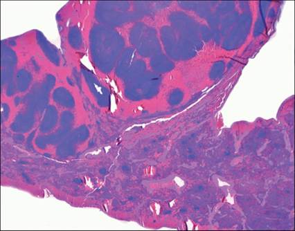
Figure 5.69 Dog, spleen, histology. Portion of spleen with marginal zone lymphoma (H&E, 50? magnification) (courtesy L. Aresu).
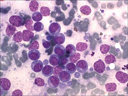
Figure 5.70 Dog, spleen, FNA, Marginal lymphoma. A single, prevalent population of medium-sized macronucleolated cells is recognizable (May–Grünwald–Giemsa, 1,000? magnification).
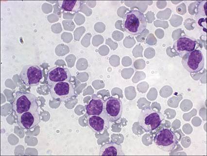
Figure 5.71 Dog, spleen, FNA. Chronic lymphocytic leukemia. A single population of mature neoplastic lymphoid cells with moderately abundant clear cytoplasm, sometimes containing fine azurophilic granules, is recognizable (May–Grünwald–Giemsa, 1,000? magnification).