Protection of the central nervous system
Meninges
The meninges (Greek for "covering") consist of three connective tissue membranes that overlay the CNS. These membranes act to cover and protect the CNS, as well as encase blood vessels and help divide gross areas of the CNS.
The three layers, named from the outermost, include the dura mater, arachnoid mater, and pia mater (Fig. 9.27). The dura mater is a tough, fibrous connective tissue layer. Surrounding the brain, it consists of two layers, with the outermost layer attached to the periosteum of the cranium. The inner layer serves as a covering over the brain and extends over the spinal cord where there is only one dural layer. Although the two dural layers over the brain are generally fused, there are several locations where the two layers separate and form dural sinuses. The dural sinuses are blood filled structures that somewhat resemble blood vessels, and which collect venous blood and return it to the internal jugular veins.
There are also areas where the dura mater extends inward from the surface of the brain forming dural folds that help stabilize gross brain structures. The largest of these folds are the following:
• Falx cerebri. This is a sickle-shaped fold of dura that extends down into the longitudinal fissure helping to separate the two cerebral hemispheres. Both the superior sagittal sinus and the inferior sagittal sinus are found within this fold.
• Tentorium cerebelli. This separates the cerebellum from the cerebral hemispheres.
• Falx cerebelli. This divides the two cerebellar hemispheres running along the vermis of the cerebellum.
The arachnoid mater (Greek for "spider") resembles a spider web and lies between the dura mater and pia mater. It forms a loose covering of the brain and never dips into the sulci or fissures. Between the arachnoid mater and pia mater is the subarachnoid space.
This space is filled with large blood vessels, as well as CSF coming from the fourth ventricle. Arachnoid granulations, or villi, project from the arachnoid, through the dura, and into the superior sagittal sinus. CSF moves by bulk flow through these granulations and into the general circulation.Finally, the pia mater closely adheres to the surface of the CNS. It contains many small blood vessels, and the pia mater adheres to these vessels for a short distance as they move into the brain.
Cerebrospinal fluid
A clear and colorless fluid, CSF has many functions: (1) It maintains a constant external environment for cells in the brain, (2) provides a route for removing harmful metabolites from the brain, (3) provides a cushion to protect the brain from trauma, (4) acts as the lymphatic system for the brain, and (5) provides a route for peptides that are released at one site and act at a distant site in the brain.
Table 9.3. Comparison of the composition of cerebrospinal fluid (CSF) and blood.
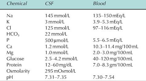
CSF is produced mostly from the choroid plexus, a tuft of capillaries found at the top of the third and fourth ventricles, as well as the floor of the lateral ventricles. The remaining CSF is formed from the ependymal cells. Although CSF originates as an ultrafiltrate of blood, its composition differs from that of plasma in several important ways. It contains less protein, calcium, and potassium, and more sodium, chloride, and hydrogen ions than plasma (Table 9.3).
Blood-brain barrier (BBB)
Neurons of the brain and spinal cord are very sensitive to alterations in their environment. As such, they are isolated from the systemic circulation by the BBB, which prevents the movement of many molecules into the CNS. The BBB is formed by the endothelial cells lining the blood capillaries. In the periphery, these cells are fenestrated; in the CNS, these cells form tight junctions.
The tight junctions are induced by factors produced by astrocytes whose end feet surround the endothelial cells of brain capillaries.To cross the BBB, a molecule either must be lipid soluble or must cross via a carrier-mediated transport system. Lipid-soluble compounds readily cross cell membranes and are thereby readily able to enter the CNS. Such compounds include carbon dioxide, oxygen, steroids, prostaglandins, and alcohol. In contrast, non-lipid-soluble compounds, such as peptides and various antibiotics, do not readily cross the BBB. However, since many essential nutrients for the brain are not lipid soluble, the BBB also possesses many carrier-mediated transport systems that allow such compounds to enter the brain at a rate far greater than that explained by their lipid solubility.
Blood-cerebrospinal fluid barrier
Although the choroid plexus is located within the ventricles, embryologically, it is derived from mesodermal tissue that is outside of the CNS. Therefore, unlike the endothelial cells in most brain capillaries, those in the choroid plexus do not have tight junctions. Instead, the ependymal cells lining the Cerebroventricles over- lying the choroid plexus have tight junctions, thus forming a barrier between the blood and CSF