Organization of the spinal cord
The spinal cord is housed in the vertebral column (Fig. 9.23). It extends from the foramen magnum at the base of the skull, to approximately the level of the first or second lumbar vertebra in an adult animal.
The spinal cord terminates as a tapered structure called the conus medullaris. It contains ascending and descending pathways to and from the brain, as well as nerve cell bodies that function in motor activity and reflexes.Table 9.2. Cranial reflexes.
| Reflex | Stimulus | Afferent Cranial Nerve | Central Synapse | Efferent Cranial Nerves | Response |
| Somatic | |||||
| Corneal reflex | Touching corneal surface | V | Motor nucleus for facial nerve | Vll | Blinking eyelids |
| Tympanic reflex | Loud noise | Vlll | Inferior colliculus | Vll | Reduced movement of auditory ossicles |
| Auditory reflex | Loud noise | Vlll | Motor nuclei of brain stem and spinal cord | III, IV, VI, VII, X, and cervical nerves | Eye and/or head movements triggered by sudden sounds |
| Vestibulo-Ocular reflex | Rotation of head | Vlll | Motor nuclei controlling eye muscles | III, IV, Vl | Opposite movement of eyes to stabilize field of vision |
| Visceral | |||||
| Direct light reflex | Light stimulating photoreceptors | Il | Superior colliculus | III | Constriction of ipsilateral pupil |
| Consensual light reflex | Light stimulating photoreceptors | Il | Superior colliculus | III | Constriction of contralateral pupil |
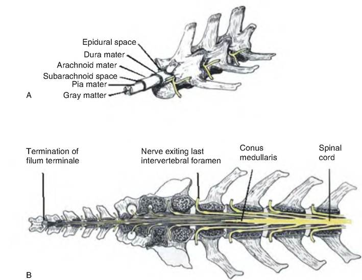
Fig.
9.23. Spinal cord. (A) The spinal cord with its various meningial layers is shown. (B) The spinal cord extends only to the level of the first or second lumbar vertebrae in adult animals where it ends in a tapered structure called the conus medullaris. A fibrous extension called the filum terminale anchors the spinal cord to the sacrum. (Modified from Getty, 1964.)Continuing as an extension of the medulla oblongata, like the brain, the spinal cord is surrounded by bone, meninges, and CSF. The dura mater over the spinal cord is a single layer that is not attached to the vertebral column. Instead, between the walls of the vertebral canal and the spinal cord is the epidural space containing loose connective tissue, blood vessels, and a layer of fat. The arachnoid mater and pia mater extend further down the spinal canal than does the spinal cord. This fibrous extension is called the filum terminale, which extends to the second sacral vertebrae and anchors the spinal cord.
In cross section, the spinal cord has a central core of gray matter consisting mostly of cell bodies and an outer region of white matter containing myelinated and unmyelinated nerve fibers (Fig. 9.24). The anterior and posterior surfaces of the spinal cord each contains a groove called the anterior median fissure and the posterior median sulcus, respectively.
The central gray area approximates the shape of a butterfly. The relative amounts of gray and white matter change at successive levels of the spinal cord. Moving from the inferior end toward the medulla oblongata, the relative amounts of gray matter decrease as white matter increases. Therefore, there are increasing numbers of fibers carrying information to the brain. The central gray area has two posterior (dorsal) horns and two anterior (ventral) horns. In addition, in the thoracic and superior lumbar regions of the spinal cord, the central gray area also has lateral horns on either side. The central gray area also has a gray commissure that connects both sides of the central gray area.
The posterior horns contain interneurons; the anterior horns contain both interneurons and cell bodies of somatic motor neurons. The lateral horns contain motor neurons of the sympathetic nervous system.The white matter of the spinal cord is grouped into three white columns, also called funiculi (long ropes). Named according to their location, they include the posterior, lateral, and anterior columns (Fig. 9.25). The
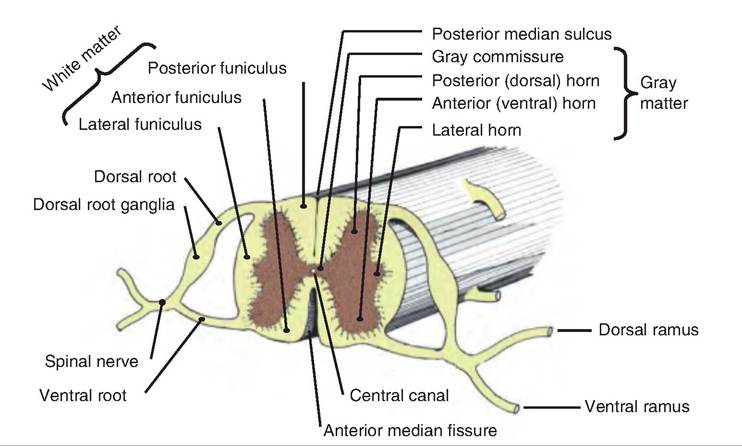
Fig. 9.24. Cross section of the spinal cord. The spinal cord has a central gray area consisting mostly of nerve cell bodies and a surrounding region of white matter consisting of fiber tracts made largely of myelinated fibers. The spinal nerves are shown on either side of the spinal cord. (Modified from Getty, 1964.)
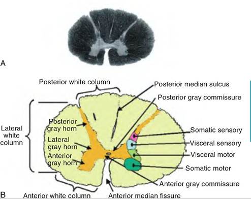
Fig. 9.25. Organization of the spinal cord. (A) A photomicrograph of a cross section of the thoracic spinal cord. (B) A drawing showing the important landmarks of the same cross section. Note that structures are bilateral.
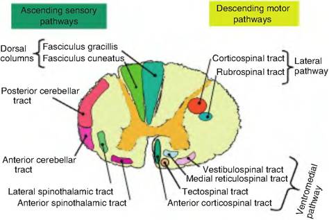
Fig. 9.26. The major ascending (sensory) and descending (motor) pathways of the spinal cord. The left side shows ascending pathways; the right side shows descending pathways within the spinal cord.
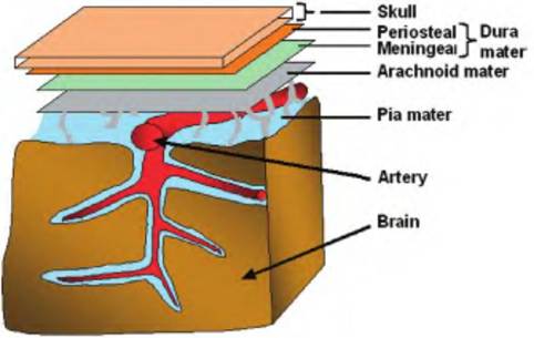
Fig. 9.27. The meninges. There are three connective tissue layers, collectively called the meninges, which cover the brain. The outermost layer, the dura mater, is a tough, fibrous connective tissue consisting of two layers: a periosteal layer attached to the inner surface of the skull and a meningeal layer. The arachnoid mater lies inside the dura mater and is a loosely knit layer. The deepest layer is the pia mater, which also courses into the brain closely adhering to capillaries as they move into the brain tissue. Between the arachnoid and pia mater is the subarachnoid space, which is filled with cerebrospinal fluid.
fiber tracts within these columns are named according to their origin and destination (Fig. 9.26).