Sample collection
Vaginal swabs are collected from the anterior aspect of the vagina to avoid contamination with either labial or cervical epithelial cells. Clean gloves should be worn. A clean cotton swab is moistened with saline.
The labia are gently separated (Figure 11.1). A sterile metal speculum can be inserted into the vagina for limited visual examination of the vagina, but this is not necessary for collection of an adequate sample (Kustritz, 2006). The moistened swab is advanced into the vagina at a 45° angle and rubbed on the vaginal surface (Figure 11.2). The swab is carefully removed, the labia are released, and the swab is gently rolled down the length of a glass slide several times (Figure 11.3). The sample is allowed to air dry and then stained with a Romanowsky-type stain (e.g. Diff-Quik®).Estrus stages (Table 11.1)
| Estrus stage | Proestrus | Estrus | Early diestrus | Late diestrus | Anestrus |
| Vaginal cytology | Squamous epithelial cells; anucleate epithelial cells; neutrophils | Anucleate epithelial cells; squamous epithelial cells | Parabasal cells; small intermediate cells; neutrophils | Small and large intermediate cells; parabasal cells; neutrophils | Parabasal cells; small intermediate cells; neutrophils |
| Uterine cytology | Endometrial cells; neutrophils; lymphocytes | Endometrial cells; neutrophils; lymphocytes | Endometrial cells; neutrophils | Degenerate endometrial cells; neutrophils; macrophages | Endometrial cells; macrophages; lymphocytes |
| Ovarian cytology | ‘Round’ cells; granulosa cells; mesenchymal cells | Granulosa cells; mesenchymal cells | Luteal cells; ‘round’ cells; granulosa cells; mesenchymal cells | Luteal cells; ‘round’ cells; granulosa cells; mesenchymal cells | Granulosa cells; mesenchymal cells |
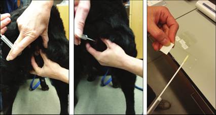
Collection of samples for vaginal cytology.
Figure 11.1 Step 1. The labia are gently separated manually to collect a vaginal swab sample.
Figure 11.2 Step 2. A moistened swab is introduced into the vagina at a 45° angle and rubbed on the vaginal surface. Care should be taken to avoid the clitoris and the labia.
Figure 11.3 Step 3. The swab is rolled onto a clean glass slide for staining and cytologic evaluation.
Proestrus
Proestrus lasts 3 days to 3 weeks in dogs and typically 1–3 days in cats. The onset of proestrus correlates with the presence of a bloody vaginal discharge, but this is often not recognized in cats. Behavioral changes including vocalizing, head rubbing, and playfulness may be noted. Maturation of ovarian follicles occurs during proestrus and anechoic follicles can be seen ultrasonographically. Cytology of a vaginal swab will contain high numbers of superficial squamous epithelial cells, low to moderate numbers of anucleate squamous epithelial cells, and low numbers of neutrophils (Figures 11.4, 11.5). Both types of epithelial cells are considered cornified epithelial cells because their cytoplasm contains keratin. These cells are large and polygonal to angular with pink or light blue to aqua colored cytoplasm. Superficial squamous epithelial cells have a small round nucleus with dense chromatin, whereas (as the name implies) no nucleus is present in anucleate squamous epithelial cells.
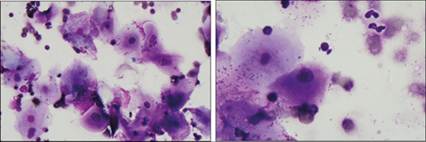
Proestrus. Vaginal swab cytology from a 2-year-old, intact female mixed-breed dog.
Figure 11.4 The majority of cells are large, cornified, nucleated squamous epithelial cells with fewer anucleate squamous epithelial cells. One, more rounded, large intermediate cell is present. Bacterial cocci and fewer rods are seen adhered to the epithelial cells. Neutrophils are expected but are not present in this image. Low numbers of erythrocytes are noted in the background (Diff-Quik®, 500? magnification).
Figure 11.5 Large, cornified, nucleated squamous epithelial cells predominate. Lower numbers of anucleate squamous epithelial cells are seen. A few nondegenerate neutrophils are present. Low numbers of erythrocytes and scattered bacterial organisms are observed in the background (Diff-Quik®, 1,000? magnification).
Estrus
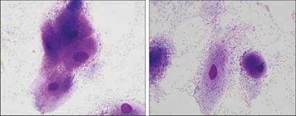
Estrus lasts 3 days to 3 weeks in dogs and cats. This stage is associated with receptiveness to mating. During this stage, estrogen concentration decreases, triggering a peak in luteinizing hormone (LH) concentration, ovulation occurs, and progesterone concentration increases sharply. In dogs, ovulation occurs 36–50 hours after the LH peak (Kustritz, 2012). Ultrasound of the ovaries shows flattening of follicles and thickening of the corpora luteal walls. Cytologically, nearly 100% of cells from a vaginal swab are cornified epithelial cells with >50% of these cells being anucleate squamous epithelial cells. Neutrophils are notably absent (Figures 11.6, 11.7).
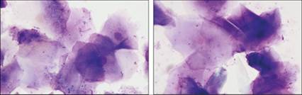
Figures 11.6,11.7 Estrus. Vaginal swab cytology from a 3-year-old, intact female Labrador Retriever. The majority of cells are cornified, anucleate, squamous epithelial cells. Low numbers of nucleated squamous epithelial cells and bacterial organisms are seen, but neutrophils are not present (Diff-Quik®, 1,000? magnification).
Diestrus
Diestrus in dogs lasts 50–80 days if not pregnant or until whelping (58–68 days) if pregnant. In cats, diestrus typically lasts 7–14 days if not pregnant or for the duration of pregnancy (63–69 days). Corpus lutea are maintained during diestrus. Decreasing LH and prolactin concentrations and an increase in prostaglandin concentration may trigger regression of the corpora lutea during the late stages of diestrus (Kustritz, 2012).
In early diestrus, vaginal cytology samples contain noncornified parabasal and small intermediate epithelial cells as well as several neutrophils (Figures 11.8, 11.9).
Parabasal cells are smaller than cornified epithelial cells and are round with a moderate amount of basophilic cytoplasm and a large round nucleus with a slightly open chromatin pattern. Intermediate cells are at an intermediate stage of maturation between parabasal cells and squamous epithelial cells. During maturation of surface epithelial cells, cellular diameter increases and nuclear diameter decreases. Therefore, small intermediate cells are round to polygonal and, compared with parabasal cells, have slightly more abundant, lightly basophilic cytoplasm and a round nucleus with moderately condensed nuclear chromatin. Large intermediate cells have still more abundant polygonal, lightly basophilic cytoplasm and have a slightly smaller nucleus with denser chromatin. In late diestrus, the number of parabasal cells decreases, small and large intermediate cells are frequent, and neutrophils begin to decrease (Figures 11.10, 11.11). Rare lymphocytes and eosinophils may be present. 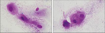
Figures 11.8,11.9 Early diestrus. Vaginal swab cytology from a 4-year-old, intact female Collie (Diff-Quik®, 1,000? magnification). Figure 11.8: Two parabasal cells (left and center) and a cell transitioning into a small intermediate cell (lower right) are present. A partially lysed, small intermediate cell also is seen (upper right). A large number of bacterial organisms are observed. Moderate numbers of neutrophils are expected but are not present in this image. Figure 11.9: Three parabasal cells are shown. There are a large number of bacterial organisms in the background. Moderate numbers of neutrophils are expected but are not present in this image.
Figures 11.10,11.11 Mid to late diestrus. Vaginal swab cytology from a 2-year-old, intact female Beagle (Diff-Quik®, 1,000? magnification).
Figure 11.10: Three cells transitioning from parabasal to small intermediate cells (top) and two large intermediate cells (bottom) are shown. There are a large number of bacterial organisms in the background. Low numbers of neutrophils are expected but are not present in this image. Figure 11.11: A large intermediate cell (left) and a parabasal cell (right) are present. A large number of bacterial organisms also are observed. Note: low numbers of neutrophils are expected but are not present in this image.
Anestrus
Anestrus lasts anywhere from 30 to 240 days in dogs (Kustritz, 2012), but may not occur at all in queens. In cats, the occurrence of anestrus is dependent on photoperiod (Faya et al., 2011). During this stage, involution of the uterus occurs. Estrogen concentration increases toward the end of anestrus causing the LH concentration to increase, which stimulates growth of new follicles in the ovary (Kustritz, 2012). Cytologically, vaginal samples taken during anestrus contain predominantly parabasal cells, moderate numbers of small intermediate cells, and low numbers of neutrophils (Figures 11.12, 11.13).

Figures 11.12,11.13 Anestrus. Vaginal swab cytology from a 1-year-old, intact female Boxer (Diff-Quik®, 1,000? magnification). Figure 11.12: A group of parabasal cells are present. A large number of bacterial organisms are also observed. Figure 11.13: One small intermediate cell (left) and two parabasal cells (right) are present. A large number of bacterial organisms also are observed. Note: low numbers of neutrophils are expected but are not depicted in these images.
Inflammation
Vaginitis or vulvovaginitis may be difficult to assess cytologically because low to moderate numbers of neutrophils are a normal finding during proestrus, diestrus, and anestrus. Additionally, during proestrus and estrus, the vulva may be mildly edematous and swollen.
Studies of vaginal bacterial flora in healthy dogs indicate that extracellular and phagocytized intracellular bacteria can be observed in vaginal cytology samples (Groppetti et al., 2012). Bacteria commonly cultured from canine vaginal swabs include Enterococcus faecalis, Streptococcus β-haemolyticus, Escherichia coli, Pasteurella multocida, and Staphylococcus spp. (Groppetti et al., 2012; Maksimovic et al., 2012). If clinical signs strongly support inflammation as a differential, and cytologic evaluation indicates suppurative or pyogranulomatous inflammation with or without intracellular bacteria, then a diagnosis of vaginitis or vulvovaginitis can be made (Figure 11.14). 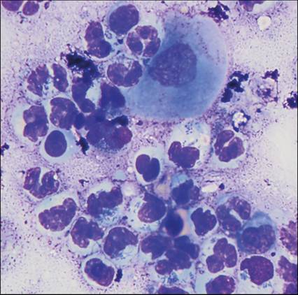
Figure 11.14 Bacterial vaginitis. Vaginal discharge cytology from a 6-year-old, spayed female Labrador Retriever mix. Clinical history included 2 years of intermittent, mucinous, bloody vaginal discharge. The cytology sample has a stippled, proteinaceous background, rare nucleated squamous epithelial cells, and large numbers of degenerate neutrophils. Many of the neutrophils contain phagocytized bacterial organisms (Wright–Giemsa, 1,500? magnification).
Neoplasia
Neoplasia of the vulva must be distinguished from an enlarged clitoris (often observed in intersex animals) or from vaginal prolapse. Neoplastic conditions of the vulva and vagina account for 2.4–3% of tumors in dogs, and approximately 83% of these tumors are benign (Thacher & Bradley, 1983; Saba & Lawrence, 2013). Leiomyoma, fibroma, and fibroleiomyoma are considered more common tumors, whereas lipoma, polyps, melanoma, myxoma, myxofibroma, and adenocarcinoma are uncommon (Ortega-Pacheco et al., 2012).
Leiomyoma/fibroleiomyoma
Benign mesenchymal tumors involving smooth muscle of the vulva, vestibule, and vagina have a similar cytologic appearance to leiomyomas arising from other areas of the body (Figures 11.15, 11.16). The overall cellularity of the sample may be scant. Cells tend to have thin, elongated, lightly basophilic, spindle-shaped cytoplasm and a centrally located, thin, long, oval nucleus with dense to stippled chromatin. Cells are generally uniform in appearance and lack overt criteria of malignancy. Histopathology is needed for a final definitive diagnosis of leiomyoma or fibroleiomyoma.
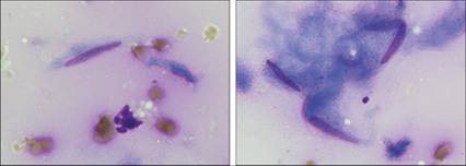
Figures 11.15,11.16 Leiomyoma (Wright–Giemsa, 1,500? magnification). Figure 11.15: A small spindle-shaped cell with a thin, long, oval nucleus is shown. Small cytoplasmic fragments, low numbers of erythrocytes, and a partially lysed neutrophil also are present. Figure 11.16: Four intact cells have spindle-shaped to wispy basophilic cytoplasm and a thin, long, oval nucleus with finely stippled chromatin and an indistinct round nucleolus. Several cytoplasmic fragments are observed. Note: differential diagnoses include leiomyoma, leiomyosarcoma, and other soft tissue sarcomas. Histopathology is needed to confirm the cytologic diagnosis.
Fibroma
Fibromas tend not to exfoliate well when aspirated for collection of a cytologic sample. These tumors are derived from fibroblasts, so cells have a moderate amount of lightly basophilic, spindle-shaped cytoplasm and a centrally located, oval nucleus. The chromatin of these cells is usually stippled and there are typically two small nucleoli, located apart from each other at opposite ends of the oval nucleus (Figure 11.17).
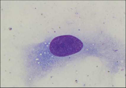
Figure 11.17 Reactive fibroblast. This cell has abundant spindloid basophilic cytoplasm and a centrally located oval nucleus with stippled chromatin and two, relatively indistinct, round nucleoli that are mildly variable in size (Wright–Giemsa, 2,000? magnification).
Polyp
Vaginal polyps are commonly observed in older dogs and tend to be solitary masses located on the ventral aspect of the vagina. These lesions cannot be distinguished from leiomyoma on gross examination and are often ulcerated (Brown et al., 2012; Foster, 2012a). In general, polyps are difficult to diagnose cytologically because cells are morphologically identical to hyperplastic epithelial cells, which are well-differentiated and may not to exfoliate well.
Adenocarcinoma
Adenocarcinoma is not a common tumor of the vulva, vestibule, or vagina. However, the lesion is worth mentioning here because the cytology and histology are similar to what is seen in samples from apocrine gland anal sac adenocarcinomas. Cytologically, these lesions have a neuroendocrine appearance with abundant lightly basophilic cytoplasm that has indistinct cell junctions and is associated with several small, rounded nuclei. Occasionally, nuclei may be loosely arranged in a circle around a central area of cytoplasm, forming acinar structures (Figure 11.18). Histologically, these tumors are classic adenocarcinomas with nests of neoplastic polygonal epithelial cells and frequent acini.
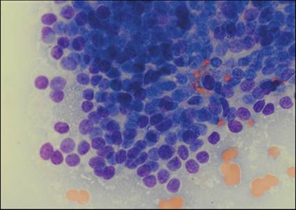
Figure 11.18 Adenocarcinoma. FNA of a clitoral mass on an 11-year-old, spayed female Basset Hound. The sample is highly cellular with several clusters of cells that have a neuroendocrine appearance. Cells have lightly basophilic cytoplasm with indistinct cytoplasmic borders and a small, round nucleus with dense chromatin. Nuclei arranged in a circular, acinar structure are observed slightly lower and to the left of the center of the image (Wright–Giemsa, 1,000? magnification).
Transmissible venereal tumor
Transmissible venereal tumors (TVTs) likely arise from canine histiocytic cells (Mozos et al., 1996; Marchal et al., 1997). Theoretically, this tumor line developed in a dog approximately 11,000 years ago (Murchison et al., 2014). Both male and female dogs can develop TVTs. The tumors tend to be present on the preputial and vaginal areas as well as around the nose. The lesion is typically a large, multilobular, ulcerated mass that exfoliates very easily by fine needle aspiration (FNA). Cytologic samples contain large numbers of round cells with abundant lightly basophilic cytoplasm; numerous clear, distinct cytoplasmic vacuoles; and a round, centrally located nucleus with finely stippled chromatin. Mitotic figures are frequently observed cytologically (Figures 11.19, 11.20). In immunocompetent animals, TVTs may spontaneously regress and are responsive to vincristine; however, metastasis is not uncommon in unhealthy animals (Foster, 2012a).
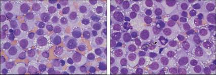
Figures 11.19,11.20 Transmissible venereal tumor. FNA of a vaginal mass from a 4-year-old, intact female mixed-breed dog. Cells exfoliate well and are individualized and round. Most cells have abundant, lightly basophilic cytoplasm with numerous, clear, distinct, round vacuoles. Nuclei are large and round with stippled chromatin and often a prominent, large, round nucleolus. Moderate anisocytosis and anisokaryosis are noted. A mitotic figure is shown at the center of Figure 11.20 (arrow). Low numbers of erythrocytes are trapped in between the neoplastic cells (Wright–Giemsa, 1,000? magnification).