Cytology of the uterus
Introduction
Uterine cytology is not commonly performed in dogs and cats. Touch imprints of the mucosal surface of the uterus or of uterine lesions may be obtained perioperatively.
Collection of cells via vaginal endoscopy with transcervical catheterization and uterine flushing has been described (Wilson, 2001). As expected, the estrous cycle greatly affects the cytologic appearance of uterine samples (Table 11.1). In dogs, during proestrus and estrus, Groppetti et al. (2010) reported finding clusters of endometrial cells with few neutrophils and rare lymphocytes. In early diestrus, they saw similar clusters of endometrial cells and few neutrophils, but lymphocytes were not observed (Figure 11.21). In late diestrus, endometrial cells became degenerate with evidence of cytoplasmic vacuolation and nuclear pyknosis, and inflammatory cells were not found. As dogs entered anestrus, endothelial cells became foamy and macrophages and neutrophils were observed. Finally, during late anestrus, endometrial cells appeared healthy and a mixed mononuclear inflammatory cell population was noted, including macrophages, lymphocytes, and plasma cells. 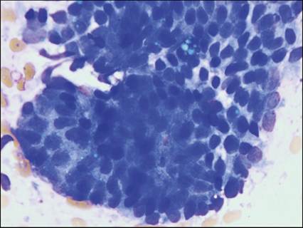
Figure 11.21 Endometrial cells. Impression smear from the uterus of an adult cat. A sheet of endometrial cells is shown. Cells have lightly basophilic, cuboidal to columnar cytoplasm and a small, round, nucleus with dense chromatin (Wright–Giemsa, 1,000? magnification).
Inflammation
Metritis is uncommon in dogs and cats, but there is a moderate amount of information about endometritis in dogs. At least 70% of canine endometritis cases are caused by bacterial infection (Fontaine et al., 2009). Bacteria commonly isolated from uterine samples include Escherichia coli, Proteus spp., Streptococcus spp., and Staphylococcus spp.
(Ortega-Pacheco et al., 2012). Cytologic sampling of a uterus affected by endometritis is unlikely to be rewarding, but may include endometrial cells, rare fibroblasts, and increased numbers of neutrophils.Pyometra and uterine stump pyometra are diagnosed more commonly in dogs and cats. Cytologic samples from uterine flushing contain degenerate endothelial cells, large numbers of neutrophils, and scattered macrophages, lymphocytes, and plasma cells (Groppetti et al., 2010). Additionally, necrotic material can be observed in vaginal smears from most of these patients (Figure 11.22).
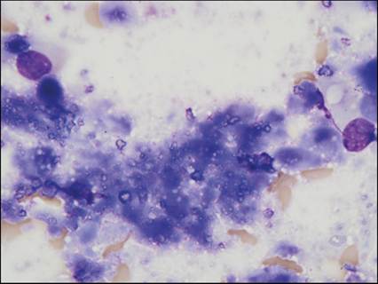
Figure 11.22 Necrosis. Blue–gray necrotic cellular debris may be observed in vaginal smears of dogs with pyometra if uterine epithelial cells are dying and sloughing off. Small, nonstaining pieces of mineralized material may be associated with the necrosis. Rare lysed nuclei are seen (Wright–Giemsa, 1,000? magnification).
Hyperplasia
Endometrial hyperplasia is uncommon in cats. In dogs, it is often cystic and has been associated with the development of pyometra (Schlafer & Gifford, 2008). In patients with cystic endometrial hyperplasia, cells collected by vaginal endoscopy with transcervical catheterization and uterine flushing included degenerate, foamy endometrial cells, and neutrophils (Groppetti et al., 2010).
Neoplasia
Leiomyoma/leiomyosarcoma
Uterine neoplasms in dogs and cats are very rare. In dogs, most uterine tumors are benign leiomyomas (Saba & Lawrence, 2013). As described for the vaginal area, the overall cellularity from a leiomyoma may be low. Smooth muscle tumor (leiomyoma and leiomyosarcoma) samples contain spindle-shaped cells with elongated, lightly basophilic cytoplasm. Benign tumors have a centrally located, thin, long, oval nucleus with dense to stippled chromatin. More malignant cells can have larger, plumper nuclei and coarse chromatin with one or more prominent nucleoli (Figure 11.23).
Other characteristics of malignancy, including binucleation, anisocytosis, and anisokaryosis, are expected with leiomyosarcomas. Neoplastic smooth muscle cells can look similar to other sarcoma cells; therefore, histopathology is needed for a final definitive diagnosis of leiomyoma or leiomyomasarcoma. 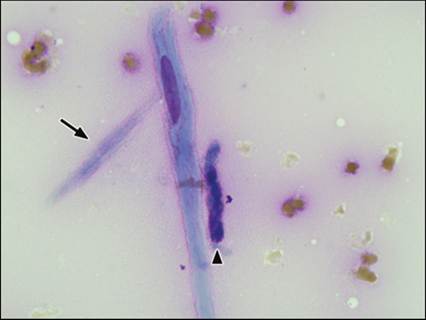
Figure 11.23 Leiomyosarcoma. The intact cell in the center of the image is strap-like with elongated rectangular basophilic cytoplasm and a thin, long, oval nucleus with finely stippled chromatin, and an indistinct round nucleolus. A cytoplasmic fragment is observed to the left of the large cells (arrow). A smaller spindle-shaped cell with a high nuclear to cytoplasmic ratio is seen at the right of the image (arrowhead). There are low numbers of erythrocytes in the background. Differential diagnoses include leiomyoma, leiomyosarcoma, and other soft tissue sarcomas. Histopathology is needed to confirm the cytologic diagnosis (Wright–Giemsa, 1,000? magnification).
Adenocarcinoma
Although rare overall, adenocarcinoma of the uterus is the most common uterine tumor in cats (Saba & Lawrence, 2013). These tumors are typically diagnosed histologically. Immunohistochemical staining indicates they are positive for cytokeratin, cyclo-oxygenase-2, E-cadherin, and β-catenin and may express progesterone receptors (Gil da Costa et al., 2009).