Cytology of ovarian tissue
Introduction
It is unusual to obtain cytology samples from ovaries of dogs and cats. Ovarian remnant syndrome is the most common complication associated with this tissue; diagnosis is typically based on the occurrence of estrous behavior in ovariectomized animals.
A vaginal cytology sample containing nearly 100% cornified epithelial cells is evidence of estrus and supports the clinical suspicion of an ovarian remnant. Cytologic samples of the ovary may be collected by ultrasound-guided FNA or perioperative impression smears. Cytology of healthy ovarian tissue varies throughout the estrous cycle. Recently, Piseddu et al. (2012) described their cytologic findings in 16 dogs undergoing elective ovariohysterectomy (Table 11.1). Low numbers of benign mesenchymal cells were seen in ovarian aspirates at all stages of the estrous cycle. Luteal cells were only observed in dogs during diestrus. These cells are large and have abundant basophilic cytoplasm, distinct cytoplasmic vacuoles, and a rounded nucleus with finely stippled chromatin (Figure 11.24). A small amount of extracellular matrix was reported in some samples during proestrus and diestrus. Ovarian cytology samples from dogs in proestrus often contained ‘round’ cells (possibly germ cells) and a few granulosa cells. During estrus, no round cells were identified but a few granulosa cells were seen. In diestrus, luteal cells were apparent in most animals and often there were moderate numbers of round cells and many granulosa cells. Finally, during anestrus, there were no round cells and a few granulosa cells. Four of the 16 dogs were sexually immature; ovarian aspirates from these animals contained moderate numbers of granulosa cells and few round cells and mesenchymal cells. Granulosa cells often exfoliate in loose clusters and have a scant to moderate amount of basophilic cytoplasm and small, round nuclei with dense chromatin (Figure 11.25). 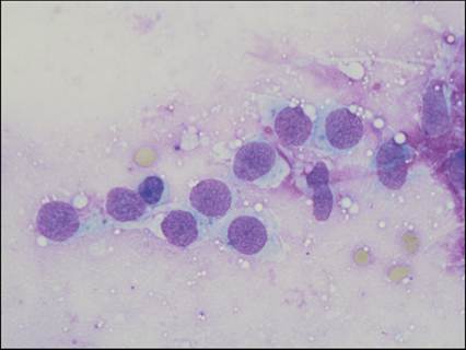
Figure 11.24 Luteal cells. FNA of an ovary from an adult cat. There are several individualized, rounded cells with lightly basophilic cytoplasm and a large, round nucleus with finely stippled chromatin. A single prominent nucleolus can be seen in some cells. One smaller granulosa cell (third cell from the left) also is present (Wright–Giemsa, 1,000? magnification).
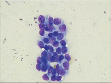
Figure 11.25 Granulosa cells. Imprint of an ovary from an adult cat. A loose sheet of granulosa cells is shown. Cells are round and have a moderate amount of lightly basophilic cytoplasm and a round nucleus with dense chromatin (Wright–Giemsa, 1,000? magnification).
Inflammation
Oophoritis is extremely uncommon in dogs and cats. Bacterial oophoritis must be differentiated from granulomas caused by feline infectious peritonitis in cats. Cytologically, one would expect to see severe neutrophilic inflammation with bacterial infection.
More on the topic Cytology of ovarian tissue:
- Barger A.M., MacNeill A.L. (Eds.). Small Animal Cytologic Diagnosis: Canine and Feline Disease. CRC Press,2024. — 536 p., 2024
- 47 Cancer of the Uterine Corpus