Cystic structures
Periovarian cysts are commonly seen in dogs and cats during ovariectomy. Intraovarian cysts may also be observed and are often derived from the rete ovarii (Foster, 2012b). Classically, cytologic samples of cystic structures are acellular and have a thin basophilic proteinaceous background consistent with the fluid aspirated from the cyst (Figure 11.26).
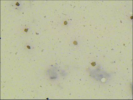
Figure 11.26 Cystic fluid. The sample is nearly acellular with a thin layer of proteinaceous material in the background of the slide. A few erythroctyes are seen (Wright–Giemsa, 800? magnification).
Neoplasia
The incidence of ovarian tumors in dogs and cats is low, with higher prevalence reported in intact animals (as expected), and ranges from 0.5 to 6.25% in dogs and 0.7 to 3.6% in cats (Saba & Lawrence, 2013). There are four categories of ovarian neoplasms: epithelial tumors and sex cord stromal tumors (specifically granulosa-theca cell tumors) are the most common tumor types, whereas germ cell tumors and mesenchymal tumors are rare.
Epithelial neoplasms
Tumors of the ovarian epithelium include undifferentiated carcinomas, papillary adenomas, adenocarcinomas, and cystadenomas, among others. Adenocarcinomas are the most common ovarian tumors in the dog and often metastasize (Patnaik & Greenlee, 1987). Carcinomas are often immunoreactive for cytokeratin, desmin, vimentin, cyclo-oxygenase-2, and endothelin-1, but nearly all carcinomas are negative for inhibin-a (Pelkey et al., 1998; Akihara et al., 2007). Most tumors are unilateral. Cysts in the contralateral ovary and/or endometrial hyperplasia may occur concurrently. Cytologically, adenocarcinomas contain clusters of rounded epithelial cells with a variable amount of deeply basophilic cytoplasm and round nucleus.
Cells occasionally may be arranged in an acinar pattern. Several characteristics of malignancy, including binucleation and prominent nucleoli, are seen.
Sex cord stromal tumors
Granulosa cell tumors are the most common ovarian sex cord stromal tumors. Most granulosa cell tumors are benign, but there is a subset (20%) that has metastatic potential (Saba & Lawrence, 2013). Immunohistochemical staining of the tumors indicates they express vimentin, S100, and often express inhibin-a and endothelin-1 (Akihara et al., 2007; Borzacchiello et al., 2010). They exfoliate well and are cytologically distinctive. Small clusters of granulosa cells are often found forming acinar structures. The cells are large and round with a moderate amount of lightly basophilic cytoplasm and an eccentric, round nucleus with stippled chromatin. Cells often contain distinct cytoplasmic vacuoles. Other, less common ovarian sex cord stromal tumors include Sertoli–Leydig tumors, luteomas, and thecomas.
Germ cell tumors
Dysgerminomas and teratomas are germ cell tumors of the ovary. Each tumor type accounts for approximately 10% of ovarian tumors in dogs (Greenlee & Patnaik, 1985). Concurrent cysts in the contralateral ovary, endometrial hyperplasia, and/or pyometra may be observed. Neither of these tumor types is commonly diagnosed cytologically. Dysgerminomas are reported to be immunohistochemically positive for vimentin and negative for cytokeratin, desmin, S100, and inhibin-a (Akihara et al., 2007). Teratomas are comprised of at least two types of embryonic tissue (ectoderm, mesoderm, and/or endoderm). Histologically, haired skin, bone, cartilage, adipose tissue, and other tissue types are often observed together in a disorganized mass (Foster, 2012b). Cytologically, teratomas are difficult to diagnose definitively. The malignant version of this tumor (teratocarcinoma) is extremely rare.