Male reproductive system
SEMEN
Semen collection and storage
Canine semen can be collected by digital massage. Having a bitch in estrus present can be beneficial. Ideally a latex collection cone is used; however, a sterile, clear, plastic bag (e.g.
a baby bottle liner) is adequate for collection of the ejaculate. Digital massage is performed by exposing the tip of the penis, placing the collection bag over the penis, and pushing the prepuce back over the bulbis glandis. Pressure is applied to the penis using a forward and backward movement. As the dog begins to thrust, the ejaculate should begin to fill the collection bag. Three fractions of ejaculate can be identified. The first fraction is clear to slightly cloudy and does not contain sperm. The second fraction should be cloudy and white and is sperm-rich. The third fraction is clear and is from the prostate. The three portions of ejaculate together have a volume of 1–30 ml (Johnston, 1991). Typically, the collection bag is removed as soon as the prostatic fraction is observed and analysis of semen is performed on the combination of the first and second fractions of the ejaculate.Collection of semen from cats can be done using a teaser queen and artificial vagina or by electroejaculation. The first method may require some training before successful collection of fluid. The total volume of the feline ejaculate ranges from 110 to 740 μl (Zambelli & Cunto, 2006).
Analysis of semen
Canine semen samples are evaluated for fluid volume, clarity, and color; aerobic and anaerobic bacteria; pH (reference interval 6.3–6.7); alkaline phosphatase concentration; and motility, quantity, and morphology of spermatozoa (Johnston, 1991). Samples from healthy dogs should have motility of spermatozoa. Greater than 70% of sperm should be progressively motile in semen. To quantitate the concentration of spermatozoa, the sample is diluted 1:106, sperm are counted using a hemocytometer, and the number of sperm/ml is calculated.
Healthy dogs produce 50–500 million spermatozoa/ml of ejaculate. Cats produce 3–600 million sperm per ejaculate sample. A cytocentrifuged sample of the fluid is stained with a Romanowsky-type stain and examined microscopically to look at the morphology of the sperm. Abnormalities in spermatozoa are described as primary or secondary defects. Fewer than 20% of sperm should have primary defects, which include proximally coiled tails, retained proximal cytoplasmic droplets, double heads, and double tails (Table 11.2). These abnormalities occur during development in the testes. Secondary defects tend to develop secondary to fever, inflammation, trauma, or infection while sperm are in the epididymis. Secondary defects include bent tails, hairpin tails, heads broken at the midpiece, retained distal cytoplasmic droplets, and detached acrosomes (Table 11.2). The percentage of sperm with secondary defects also should be alt=fig11.30.jpg>Figure 11.30 Early spermatids. Imprint of a testicle from an adult dog. Spermatids are present in large numbers. They are round cells with a moderate amount of basophilic cytoplasm that contains low to moderate numbers of distinct, clear, cytoplasmic vacuoles. The nucleus is rounded with finely stippled chromatin. Binucleated and multinucleated cells are frequently seen (Wright–Giemsa, 1,000? magnification).
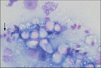
Figure 11.31 Cap phase spermatid. Imprint of a testicle from an adult dog. One cap phase spermatid is shown at the left of the image (arrow). The cell has a nucleus with a rounded edge and a pointed edge. Several early and late spermatids are observed. A few Sertoli cells and a spermatocyte also are present (Wright–Giemsa, 1,000? magnification).
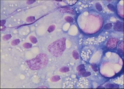
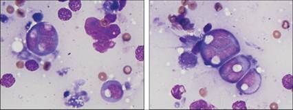
Figure 11.32 Late spermatids. Imprint of a testicle from an adult dog.
Several late spermatids are present in this image. Cell cytoplasm is scant and pale. The nucleus is dense with an oval edge and a flattened edge (Wright–Giemsa, 2,000? magnification).
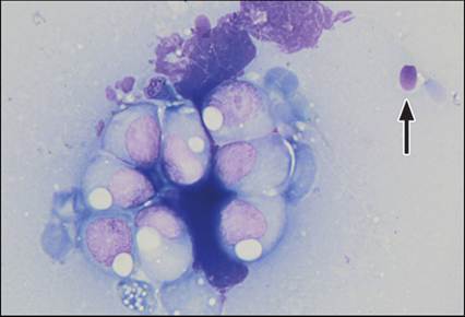
Figure 11.33 Spermatozoon. Imprint of a testicle from an adult dog. A mature spermatozoon is shown at the upper right of the image (arrow). The cell has a pale, light blue head and a long, nonstaining tail. Several early spermatids and two late spermatids also are shown (Wright–Giemsa, 1,500? magnification).
Infertility
Morphologic abnormalities observed in spermatozoa in wet mounts of semen samples can also be recognized during cytologic evaluation of testicular aspirates. These abnormalities are important findings in infertile animals.
Inflammation
Neutrophils can be seen cytologically in testicular aspirates from animals affected by orchitis, periorchitis, or epididymitis. In these cases, it is recommended that the clinician submit a sample for bacterial culture for detection of Babesia canis. Transient, sterile, neutrophilic inflammation is observed following chemical castration by injection of the testes with zinc compounds. Inflammation should be minimal and decreased semen volume and azoospermia are expected by 3 months following this procedure (Fahim et al., 1993; Fagundes et al., 2014).
Hematocele and hydrocele
Hematoceles and hydroceles are formed when blood or ascitic fluid, respectively, fills the cavity formed by the vaginal tunics. Aspiration of these lesions will yield a poorly cellular fluid sample that may contain low numbers of macrophages. If the macrophages contain phagocytized erythrocytes and black pigment (hemosiderin), a hematocele is suspected.
Neoplasia
Testicular tumors include epithelial tumors, sex cord stromal tumors, and germ cell tumors. In dogs, testicular tumors tend to be benign (metastasis occurs in Interstitial cell tumor.
FNA of a testicular mass on an adult intact male Rottweiler. Figure 11.41: Cells are aggregated and have a small amount of lightly basophilic cytoplasm and a small, round nucleus with dense chromatin (Wright–Giemsa, 1,000? magnification). Figure 11.42: A small prominent nucleolus can be appreciated in some of the partially lysed cells in Figure 11.40 (Wright–Giemsa, 1,500? magnification).
Germ cell tumors
Seminomas are common germ cell tumors of dogs. As with Sertoli cell tumors, seminomas more often affect cryptorchid testicles. Grossly, seminomas are soft, small to lobulated, and ivory. Aspirates of seminomas contain large, individualized cells with a moderate amount of basophilic cytoplasm and a large, round nucleus that has finely stippled chromatin. A prominent nucleolus may be observed. Binucleation and mitotic figures are frequent findings (Figures 11.43, 11.44).
Figures 11.43,11.44 Seminoma. FNA of a testicular mass on a 14-year-old, intact male Cocker Spaniel. Large, individualized, round cells with basophilic cytoplasm and low numbers of round, clear, distinct cytoplasmic vacuoles are observed. These cells have one or more round to oval nuclei with stippled chromatin. Several lysed cells and few erythrocytes are seen in the background (Wright–Giemsa, 1,500? magnification).