Signalment and History
Nasopharyngeal or inflammatory polyps originate from the eustachian tube or from the middle ear mucosa. Most of these polyps have their origin in the middle ear; however, when they originate in the eustachian tube, growth is usually directed toward the throat.
Nasopharyngeal polyps can be found exiting the eustachian tube on the lateral wall of the oropharynx, beneath the soft palate. Retraction of the soft palate with a spay hook reveals their presence. The cat with an oropharyngeal location of its polyp shows respiratory symptoms such as stertorous respiration, voice changes, wheezing, dyspnea, and dysphagia.1The predominant clinical signs in a cat with an inflammatory polyp of the ear canal are discharge from the ear canal and head shaking and/or head tilt. Polyps are rarely bilateral. Nystagmus and vestibular disease may be present in severe cases of aural polyps, because pressure on the round and oval windows from the enlarging mass increases pressure in the endolymph within the semicircular canals.
Several presentations can be seen when a cat has a polyp in the ear canal. Some cats with an aural polyp are presented to the veterinarian with a waxy accumulation deep in the ear canal (Figure 16-1). This may look similar to a ceruminolith or wax plug. The polyp mass cannot be visualized because it is under the wax.
Some cats have dried, crusty material in the ear, similar to the discharge seen in Otodectes infections. The middle ear mucosa is a respiratory epithelium capable of producing copious amounts of mucus when it is inflamed. The mucus and pus that leak from the bulla into the external ear canal can dry out and become flaky, giving the impression that the cat has ear mites. More commonly, cats with polyps have a copious mucopurulent discharge in the affected ear due to liquid exudates filling the ear canal (Figure 16-2).
After cleaning the cat’s ear, otoscopic examination reveals a pink to red, fleshy, mobile mass deep in the ear canal (Figures 16-3 and 16-4).
When the mass is visible in the external ear canal, it has already protruded through the eardrum, having ruptured it as the polyp grew outward from the tympanic bulla. When this happens, a secondary otitis media can develop as bacteria gain access to the middle ear mucosa.Manipulating the polyp mass often causes the release of material from the bulla into the external ear canal because the polyp acts as a seal for these middle
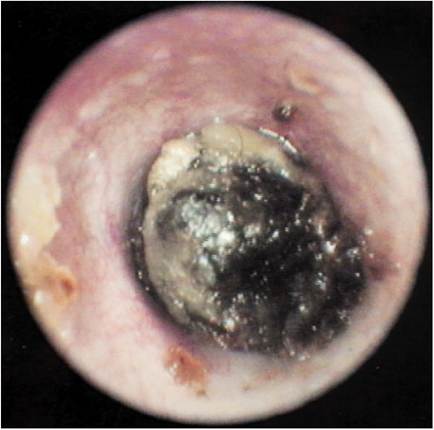
Figure 16-1
Cat with a waxy accumulation in the ear canal. This mass was located deep in the horizontal portion of the cat's ear canal. The cat was presented for a head tilt and ataxia.
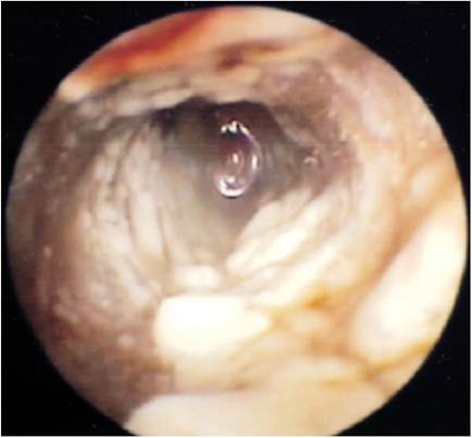
Figure 16-2
Liquid mucus and pus filling the ear canal of a kitten. After flushing the ear canal, the polyp was revealed (see Figure 16-4).
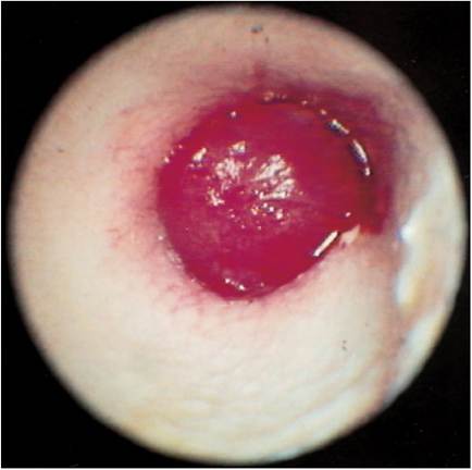
Figure 16-3
Nasopharyngeal polyp occluding the ear canal of a cat. The waxy accumulation in Figure 16-1 was removed to reveal this red, fleshy mass in the cat's ear canal.
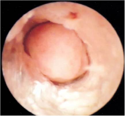
Figure 16-4
After the mucus and pus were removed from the ear canal from the cat in Figure 16-2, this fleshy mass was evident and could be pushed back into the middle ear. No eardrum is present in cats with polyps.
ear exudates. Polyps can become quite large and may reach the diameter of the ear canal, effectively forming a plug that seals in the secretions in the bulla under it.