Microbiology
Many kittens and grown cats have unrecognized middle ear disease often as a sequela to upper respiratory disease. Otitis media is a consistent feature when nasopharyngeal polyps are found in the ear canal.
Many bacterial pathogens have been cultured from the middle ear after bulla osteotomy including Pasteurella, streptococci, staphylococci, and occasionally Bacteroides and Pseudomonas.2 Some bacteria isolated from the middle ear in cats with polyps originate from the upper respiratory mucosa, and some pathogens originate from the external ear canal epithelium. Routine aerobic cultures for respiratory pathogens rarely include Mycoplasma, Bordetella, or Chlamydia, which may be involved in respiratory disease and middle ear disease of cats.3 It was once thought that viruses played a role in development of polyps. In one study, tissues from inflammatory polyps were assayed for feline calicivirus and feline herpesvirus-1 by polymerase chain reaction (PCR). Failure to detect either of these viruses suggests that the persistence of these viruses is not associated with the development of inflammatory polyps.4 In another study, polyps were induced in rats by placing type 3 pneumococci into one middle ear cavity of each rat. In 44% of the rats a polyp developed in the experimentally infected ear. None of the rats developed a polyp in the untreated ear.The sequestered inflammation and infection lead to changes in the middle ear mucosa. It is theorized that the chronic irritation of the middle ear mucosa leads to the formation of polyps. Initially there is a rupture in the respiratory epithelium lining the tympanic bulla or the eustachian tube, followed by intraluminal protrusion of the lamina propria through the epithelial defect. Then the respiratory epithelium covers this protrusion. As the epithelium covering the polyp becomes damaged by infection or by the physical trauma of its own movement against the surrounding tissue, this process repeats a number of times, resulting in significant production of fibrous stroma and enlargement of the mass.
Often the mass becomes lobulated (Figure 16-5).Grossly, a nasopharyngeal polyp is pedunculated, having a narrow stalk at its origin and becoming a large fleshy mass as it grows. Histologically, a polyp is a hyperplastic inflammatory proliferation of the middle ear mucosa. A polyp is composed of well-vascularized fibrous tissue stroma covered with a respiratory epithelium, which is often ulcerated. The stroma is edematous, and the submucosa contains a mixed population of acute and chronic inflammatory cells, including neutrophils, macrophages, and plasma cells. Variable amounts of lymphocytes may also be present. It is difficult to determine the exact point of origin of a polyp histologically because the respiratory epithelium of the mucous membrane in the tympanic cavity, auditory tube, and nasopharynx is continuous.
Depending on their growth pattern, polyps can grow through the auditory tube toward the nasopharnx or they may grow through the tympanic membrane. When found in the external ear canal, the enlarging polyp mass has grown through the
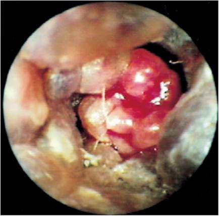
Figure 16-5
Multilobulated polyp in a cat. This type of polyp is usually more vascular than solitary-lobed polyps.
tympanic membrane, creating a permanent opening from the external ear canal to the middle ear. The middle ears of these cats have copious mucus and pus.
More on the topic Microbiology:
- Other Mycobacterial Infections in Livestock and Wildlife in Tanzania
- Zoonotic Tuberculosis in Zambia
- AVIAN CHOLERA
- Alexander’s Invasions
- 3 SELECTED SOCIO-ECONOMICALLY IMPORTANT WILDLIFE RELATED PATHOGENS AND DISEASES IN EUROPE
- CHAPTER 41 HARMFUL ALGAL BLOOMS INCLUDING Cyanobacterial toxicosis
- Limitations and Opportunities for the Use of Molecular Epidemiological Tools in Africa
- Conclusion and Recommendations