Specific joints
Intervertebral articulations
The intervertebral articulations consist of cartilaginous and synovial joints. The cartilaginous joints are formed by the intervertebral discs joining the bodies of the vertebrae.
The synovial joints are formed by caudal and cranial articular processes of the adjacent vertebrae.The first two joints within the vertebral column are atypical. The first, the atlanto-occipital joint, is a modified hinge type of the synovial joint between the occipital condyles and the cranial articular surfaces of the atlas (i.e., first vertebral vertebra). Thisjoint has a spacious joint capsule and is specialized to allow a "yes" motion. The atlanto-axial joint is a pivot type of synovial joint. It is between the dens of the axis and the cranial articulation surfaces on the atlas.
Costovertebral joints
There are two types of articulations between the ribs and the vertebral column. The head of each rib forms a ball-and-socket type of Synovialjoint, with the causal and ostal facets of adjacent vertebrae. The tubercle of each rib forms a plane type of synovial joint with the transverse process of the corresponding rib (Box 6.1).
Sternocostal joints
There is a pivot type of synovial joint between the first eight costal cartilages and the sternum. Each joint has a joint capsule and ligaments.
Costochondral joints
There is a fibrous joint between the ribs and costal cartilage. These have no synovial cavities or joint capsule.
Box 6.1 Rupture of an intervertebral disc
The rupture or degeneration of a disc between the vertebrae allows the pulpy nucleus to bulge or leak out of the disc. This usually occurs dorsally or dor- Solaterally This can result in pressure being placed on the spinal cord or spinal nerves. It most commonly occurs at the thoracolumbar junction or neck region.
Thoracic limb
Shoulder joint
Also called the glenohumeral or scapulohumeral joint, the shoulder joint is a ball-and-socket type of synovial joint.
The head of the humerus articulates with the glenoid cavity of the scapula. It contains a loose joint capsule with no true collateral ligaments. Instead, the muscles crossing the joint provide the support to minimize shoulder luxation (i.e., separation). Functionally, this is a freely movable joint (Fig. 6.35).The intertubercular, or bicipital, sulcus is a groove between the greater and lesser tubercles. This site holds the biceps brachii tendon. There is a synovial sheath around this tendon as it passes through the intertubercular groove in carnivores, pigs, and sheep. In horses, oxen, and goats, there is an intertubercular bursa found between the intertubercular groove and the bicipital tendon. The transverse humeral ligament attaches between the greater and lesser tubercles holding the biceps tendon in the intertubercular groove.
Elbow joint
The humeroradioulnar articulation is a hinged type of synovial joint allowing flexion and extension. It is a compound joint since it consists of three bones. There is a joint capsule encasing all three bones.
The humeral condyle consisting of the capitulum and trochlea articulates with the head of the radius, and the anconeal process of the ulna fits into the olecranon fossa of the humerus. The medial and lateral collateral ligaments located on the sides of the joint restrict the movement to flexion and extension.
In horses and ruminants the proximal and distal joint between the radius and ulna is fused. In carnivores, these joints are not fused. This allows some rotation of the radius and hence, some degree of supination of the forepaw, as well as return to pronation.
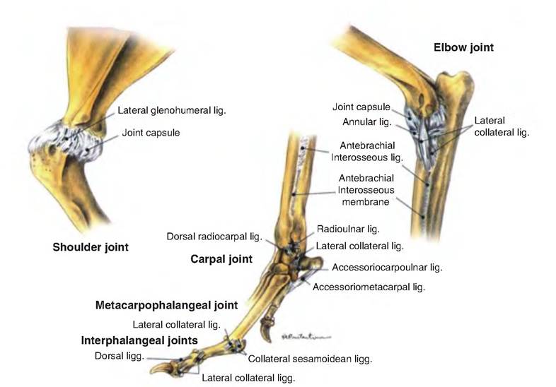
Fig. 6.35. Joints of the Thoracic Limb. The lateral aspect of the thoracic limb of the clog. (Reprinted from Constantinescu, 2002. Used by permission of the publisher.)
Carpal joint
The carpal joint consists of three main joints including the antebrachiocarpal, middle carpal, and carpometacarpal joint.
The carpal joint is a hinged type of synovial joint. The antebrachiocarpal joint consists of an articulation between the distal radius and ulna and the proximal row of carpal bones. The distal row of carpal bones articulates with the metacarpal bones, constituting the carpometacarpal joint. The middle carpal joint is between the two rows of carpal bones. There are plane joints between individual carpal bones.Pelvis
Pelvic symphysis
This is a slightly movable fibrocartilaginous joint between the hip bones (os coxae). The front portion of this joint is formed by the pubic symphysis between the two pubic bones; the caudal portion is formed by the ischial symphysis between the two ischial bones.
Sacroiliac joint
The sacroiliac joint is a relatively immobile joint between the wings of the sacrum and the ilium. It is a combination of a cartilaginous and synovial joint. Fibrocartilage unites the ilium with the wing of the sacrum.
The Sacrotuberous ligament connects the sacrum and first caudal vertebrae with the ischiatic tuberosity. This ligament stabilizes the caudal end of the sacrum between the os coxae. It is absent in cats.
Hip joint
Also called the coxal or Coxofemoral articulation, this is a ball-and-socket synovial joint between the head of the femur and the acetabulum of the hip bone. It is a freely movable (diarthrodial) joint allowing universal movement (i.e., flexion, extension, abduction, adduction, lateral rotation, and circumduction). It has no collateral ligaments; instead, its stability depends on the ligament of the head of the femur, a strong joint capsule, and a large muscle mass surrounding it. The ligament of the head of the femur connects from the acetabular cavity to the notch on the fovea capitis, on the head of the femur. Found only in horses, the accessory ligament of the head of the femur extends from the prepubic tendon through the acetabular notch under the transverse acetabular ligament to the fovea capitis of the head of the femur.
This ligament makes it harder for the horse to kick to the side, that is, cow kick, although it doesn't totally prevent it (Box 6.2).Box 6.2 Hip dysplasia
Hip dysplasia involves a malformed hip joint resulting in a progressive degenerative disease. This disease has a high incidence in some breeds of dogs. Diagnosed radiographically, the condition causes pain. Treatments include cutting the pectine- ous muscle, removing the neck and head of the femur (head and neck osteotomy), or remodeling the acetabulum by cutting the hip bones and repositioning them.
Pelvic limb
Knee (stifle joint)
The knee, also known as the stifle joint, is a compound joint involving the femur, patella, and tibia. It is a hinge type of synovial joint allowing flexion and extension with little rotation (Fig. 6.36).
The joint between the patella and femur is called the femoropatellar joint and contains a large joint capsule. The patellar ligament runs between the patella and the tibial tuberosity. Remember that the patella is a sesamoid bone, meaning that it is found within a tendon. Carnivores, pigs, and small ruminants have one patellar ligament; horses and oxen have three, including the lateral, middle, and medial.
The Iemorotibialjoint is the articulation between the femur condyles and the tibia, and has interposed menisci. These menisci include the medial and lateral menisci that sit between the tibial and femoral condyles.
The medial collateral ligament fuses with the joint capsule and medial meniscus and stabilizes the medial side of the stifle. The lateral collateral ligaments connect the lateral epicondyle and head of the fibula. It is separated from the lateral meniscus by the tendon of the popliteus muscle.
The cranial cruciate ligament originates on the cau- dolateral femur and inserts Cranially on the tibia. It prevents cranial movement of the tibia relative to the femur. The caudal cruciate ligament arises from the Craniomedial distal femur and inserts on the tibia. It prevents caudal movement of the tibia relative to the femur.
Tarsus
The tarsus, or hock, is a compound hinge type of synovial joint. It allows only flexion and extension. The Iibiotarsal portion of the tarsus is the most movable joint, and is an articulation between the proximal row of tarsal bones (i.e., the talus and calcaneus) and the fibula and tibia. The cochlea of the tibia receives the trochlear ridges of the talus. The proximal intertarsal joint is the articulation between the proximal row of tarsal bones and the central and fourth tarsal bones. The distal intertarsal joint includes the articulation between the central tarsal and tarsal bones I, II, and III.
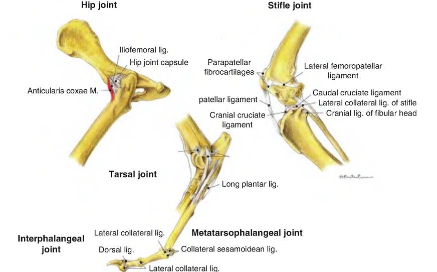
Fig. 6.36. Joints of the pelvic limb. The lateral aspect of the pelvic limb of the dog. (Reprinted from Constantinescu, 2002. With permission from the publisher.)