Joints
Types of joints
Arthrology is the study of joints. Joints are necessary to allow for the movement of the skeleton. By their structure, joints can be classified several ways according to (1) the number of articulating bones, (2) structural classification, and (3) functional classification:
1.
Number of articulating bones. A simple joint has only two articulating bones forming the joint, whereas a compound joint has three or more articulating bones.2. Structural classification. Joints can be classified by the medium holding the joint together (Table 6.4):
a. Fibrous joints. Fibrous tissue between bones holds the bones together but allows little or no movement; thus, no joint capsule is present (Fig. 6.32). These joints usually ossify later. According to details of formation, fibrous joints are classified as sutures, syndesmoses, or gomphoses.
b. Cartilaginous joints. Fibrocartilage, hyaline cartilage, or both hold the joint together (Fig. 6.33). Thesejoints only allow slight movement, so like fibrous joints, they lack a joint capsule. Two types of cartilaginous joints are synchondroses and symphyses. The best examples of synchondroses are the epiphyseal plates in long bones, which eventually close, and the joint between the first rib and the manubrium. An example of a symphysis is the pubic symphysis.
c. Synovial joints. The articular surfaces of two hyaline cartilage-covered bones are joined by a synovial joint capsule, and are freely
| Table 6.4. Classification of joints. | ||||
| Structural Class | Characteristics | Type | Mobility | Example |
| Fibrous | End of bones united | 1. Sutures | Immobile (synarthrosis) | Bones of the cranium |
| by fibrous tissue | 2. Syndesmosis | Slightly mobile (amphiarthrosis) and immobile | Distal Iibiofibularjoint | |
| 3. Gomphosis | Immobile | Articulation of a tooth with its socket | ||
| Cartilaginous | End of bones united by cartilage | 1. Synchondrosis (hyaline cartilage) | Immobile | Epiphyseal plates |
| 2. Symphysis (AbrocartiIage) | Slightly movable | Pubic symphysis | ||
| Synovial | Ends of bones covered with articular cartilage, and a joint cavity enclosed with a joint capsule | 1. Ball-and-socket 2. Pivot | Freely movable Rounded end of one bone projected into sleeve or ring on another bone; freely movable but allows only uniaxial rotation | Coxofemoral (hip) joint and glenohumeral (shoulder) joint Between atlas and dens of axis; proximal radioulnar joint in animals where pronation and supination possible |
| 3. Ellipsoidal | Both articulating surfaces are oval; freely movable allowing flexion, extension, abduction, adduction, and circumduction | Radiocarpal joints | ||
| 4. Saddle | Each articulating surface has both concave and convex areas, resembling a saddle; freely movable | Caprometacarpal joint of thumb in man | ||
| 5. Plane (or gliding) | Articulating surfaces -flat; freely movable, but only slipping or gliding motions | Intercarpal and intertarsal joints; vertebral processes | ||
| 6. Hinge | Cylindrical projection of one bone into trough-like depression of another | Knee, elbow, and interphalangeal joints | ||
movable. The structure and types of synovial joints are discussed later.
3. The functional classification of joints indicates the degree of mobility in the joint:
a. Synarthrotic. Movements in these joints are absent or extremely limited. Examples of these joints include the sutures in the cranium.
b. Amphiarthrotic. There is slight movement in these joints. Examples include the intervertebral joints of sternoclavicular joints.
c. Diarthrotic. Also called synovial joints, these joints have considerable movement. They allow for one-, two-, or three-dimensional movement, and contain articular cartilage and synovial membranes. Many such joints also contain bursae sacs. Examples include shoulder, knee, wrist, and elbow.
Synovial joints
Anatomy of synovial joints
The synovial joint is a complicated joint involving many parts. It is movable, and consists of a joint cavity, articular cartilage, joint capsule with an inner synovial membrane, and an outer fibrous layer (Fig. 6.34). The fibrous layer attaches to the periosteum on or near the articular cartilage. The synovial membrane is highly vascular, well innervated, and produces synovial fluid. Synovial fluid is viscous and acts to lubricate the joint, provide nutrients, and remove waste from the hyaline articular cartilage.
The articular cartilage is a translucent, bluish-tinged cartilage, usually hyaline, that covers the articulating surfaces of the bone. The joint cavity is unique to synovial joints and contains a trace amount of synovial fluid. Outside the fibrous layer of the joint capsule may be ligaments that hold together the bones of the
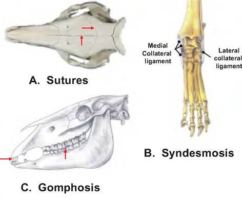
Fig. 6.32. Fib rous joints. Examples of fibrous joints include the following: (A) Sutures as found between the bones in the skull.
(B) Syndesmosis joints in which ligaments connect the bones.
(C) Gomphosis joints exemplified by the teeth located in alveolar sockets. Used with permission from Constantinescu, G.M., 2002. Clinical Anatomy for Small Animal Practitioners. Iowa State Press, Ames, Iowa.
joints. The ligaments consist of bands of white fibrous connective tissue holding the joints together.
The meniscus or articular menisci is Abrocartilage that partially or completely divides a joint cavity. Menisci are found only in the stifle and temporomandibular joints. They serve to make the joint more stable by improving the fit between two articulating bones.
A bursa is a saclike structure between different tissues that acts as a ball bearing, reducing the friction between the bones. The bursa is a flattened sac lined with a synovial membrane and contains a small amount of synovial fluid. While technically not part of the synovial joint, bursae are associated with such
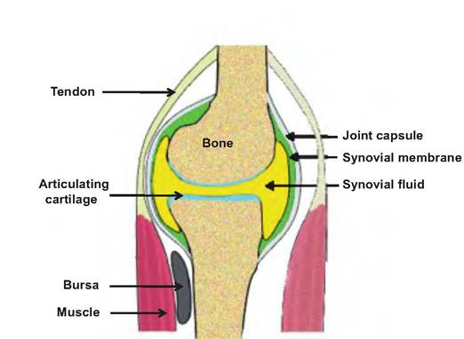
Fig. 6.34. General structure of a synovial joint. Modified from http://www.studyblue.eom/notes/note/n/ibhs-3-lecture-3/deck/4036345.
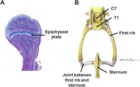
Fig. 6.33. Cartilaginous joints. (A) The epiphyseal plate located in a growing long bone. (B) The sternocostal joint located between the first rib and sternum. Used with permission from Constantinescu, G.M., 2002. Clinical Anatomy for Small Animal Practitioners. Iowa State Press, Ames, Iowa.
joints where ligaments, muscles, skin, tendons, or bones rub together. A bunion is an enlarged bursa at the base of the big toe in humans.
A tendon synovial sheath wraps completely around a tendon. It acts similar to a bursa, reducing friction between the tendons and bones.
Classification of synovial joints
The types Ofsynovialjoints can be classified as follows:
1. Ball-and-socket.
Alsocalledaspheroidortriaxial, this joint allows all movements, thus allowing the greatest range of motion. Examples include the iliofemoral (hip) joint and glenohumeral (shoulder) joint.2. Hinge. Alsocalledaginglymusormonaxialjoint, movement is limited to flexion and extension. Examples include the knee, elbow, and interpha- Iangeal joints.
3. Pivot. Also called a trochoid or monaxial joint, it allows movement limited to rotation. Examples include the atlantoaxial or proximal radioulnar joint.
4. Ellipsoidal or condyloid. Also called a condyloid or biaxial joint, it is essentially a reduced ball-and- socket joint. Ellipsoidal joints allow all angular motions, including flexion, extension, abduction, and adduction, but not rotation. Examples include the radiocarpal joints.
5. Saddle. Also known as sellar or biaxial, allows all movements except rotation. An example includes the carpometacarpal joint of thumb.
6. Plane. Also called an arthrodia, gliding, or biaxial joint, allows gliding in flexion, extension, abduction, and adduction. Such joints are present in inIercarpal and intertarsal joints.
Movements of synovial joints
Synovial joints can make various types of movements and display different ranges of motion. The range of motion of synovial joints varies from nonaxial movement, which includes slipping motions only; to uniaxial movement, involving motion in one plane; to biaxial movement (movement in two planes); and to multiaxial movement, involving movement in three planes.
There are three general types of movements possible in synovial joints: rotation, gliding, and angular. These are listed in Table 6.5.
In addition, there are special movements unique to synovial joints. The manus (hand) can undergo supination, palm-up position, and pronation, palm-down or back position. Supination involves the lateral rotation of the radius; pronation involves the medial rotation of the radius relative to the ulna. During pronation, the distal end of the radius crosses over the ulna so that the bones form an "X."
Table 6.5.
Movements within synovial joints.| Movement | Description | Example |
| Rotation | ||
| Rotation | Turning a bone around its own long axis | Femur can rotate away from median plane (lateral |
| rotation) or toward median plane (medial rotation) | ||
| NonAnguIar Movements | ||
| Gliding | One flat or nearly flat bone surface slips over | Intercarpal and intertarsal joint movements |
| another similar surface | ||
| Angular Movements | ||
| Flexion | Decreasing the angle of the joint | The elbow joint (humerus-radius/ulna) |
| Extension | Increasing the angle of the joint | The elbow joint (humerus-radius/ulna) |
| Dorsal and ventral flexion | Bending the spinal column dorsally or ventrally | The spine |
| Abduction | Moving a part away from the median plane | The shoulder joint (humerus-glenoid fossa) |
| Adduction | Moving a part toward the median plane | The shoulder joint (humerus-glenoid fossa) |
| Circumduction | Movement that traces a cone shape, thus combining | Movement of a limb in a circular motion with the |
| flexion, abduction, extension and adduction | shoulder or hip remaining essentially stationary | |
| Rotation | Movement around the long axis of a part | Radio-ulnar joint |
| Universal | All of the above movement | The shoulder joint |
Inversion and eversion are terms describing the movement of the foot. During inversion, the sole of the foot turns medially; during eversion, the sole faces laterally.
Protraction and retraction involve nonangular anterior or posterior movement along a transverse plane. When the mandible is pushed outward from the jaw, this is protraction; pulling the mandible back is called retraction.
Elevation and depression are terms used to describe shoulders or jaw movement. When the shoulders are moved dorsally, it is called elevation; lowering the shoulders is called depression. The mandible is elevated or depressed during chewing.