Structures of the integument
The integumentary system is the largest organ of the body and is an extremely important barrier to systemic injury and infection. It plays a key role in water and electrolyte homeostasis, temperature regulation, and production of vitamin D3, and acts as a sensitive sensory organ.
It is composed of the epidermis, dermis, and hypodermis (subcutis) (Figure 4.1a). Specific areas of skin are specialized and may or may not contain hair follicles and glands. Numerous vascular, neural, and lymphatic structures are intimately associated with all components of the integument. 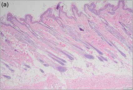
Figure 4.1a Integument histopathology. Section of skin from a 2-month-old Husky. The integument is composed of the epidermis, dermis, and subcutis. The epidermis is the most external layer (top; Figure 4.1b). The dermis lies just beneath the epidermis and is composed of collagen and elastin fibers, vasculature, and adnexal structures (including hair follicles and sebaceous glands, which are in longitudinal section in this specimen). The subcutis is composed of adipose tissue (round clear spaces) and collagen (bottom) (H&E, 50? magnification).
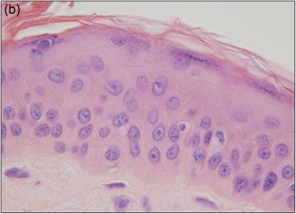
Figure 4.1b Epidermis histopathology. Section of skin from a 15-year-old domestic longhair cat. The pale cells at the bottom of the image make up the superficial dermis. Basal epithelial cells are the first layer of eosinophilic cells with large round nuclei just above the dermis. The next layer of nuclei in the middle of the eosinophilic cells is associated with the stratum spinosum. The stratum granulosum is the layer of cells that have very pale nuclei or have lost their nuclei. Some cells of the stratum granulosum just underneath the surface layer of keratinized cells contain basophilic granules.
The stratum corneum is the bright pink, flaky, keratinized material at the top of the image (H&E, 1,000? magnification).
Epidermis
Epidermal cells comprise the outermost layer of the integument, which develops from the embryonic ectoderm. Most cells of the integument are stratified squamous epithelial cells, although simple cuboidal epithelial cells form the secretory and ductal components of glands. The distinct stratified layers of the integument are derived from the basal cell layer of the epidermis (Figure 4.1b). Basal cells and epidermal stem cells are present in the stratum basale at the deepest layer of the epidermis. They are attached to a basement membrane at the interface with the dermis. Melanocytes are also found in this layer, and in the outer root sheath of hair follicles, and produce melanin which gives color to skin and hair. Basal cells are small, cuboidal cells with very distinct cell junctions and a small round nucleus. As basal cells migrate toward the most superficial layer of the epidermis, they flatten and form the cells of the stratum spinosum. These cells are larger, have more cytoplasm, and are polyhedral in shape (Welle & Linder, 2021). These cells continue to migrate toward the surface. In the stratum granulosum, cells become layered upon each other and begin to produce keratohyalin granules (keratin precursors). Their nuclei begin to shrink and become pyknotic. In some specialized areas of the skin (nose, foot pads, teats), the next layer of skin (stratum ludicum) contains enough layers of cells that it can be visualized histologically. Cells of the stratum lucidum are pale, flattened cells that often lack a nucleus. Finally, the most superficial layer of cells forms the stratum corneum, which is made up of dead, keratinized, squamous epithelial cells that eventually slough off as the skin regenerates. These cells are typically polygonal with abundant keratinized cytoplasm and have a pyknotic nucleus or lack a nucleus altogether.
Cytologically, keratin in cells is a light blue–green color and may have a glassy appearance when stained with Romanowsky-type stains. In most areas of the integument, the squamous epithelial cells at the surface become cornified and lack a nucleus. These cells are described as keratin flakes or keratinized or keratinaceous debris in cytology samples and are more angular and blue–green to pink–purple in color (Figures 4.2). 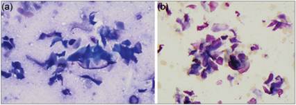
Figures 4.2a,b Superficial keratinocytes from the skin surface are large and angular or polygonal cells that stain somewhat variably with Wright–Giemsa stain. Cells may be aqua-blue to bright blue (a, 500? magnification) or eosinophilic to purple (b, 500? magnification). Importantly, superficial keratinocytes lack a nucleus.
Dermis and adnexal structures
The dermis is made up of mesenchymal cells and collagenous fibers derived from the embryonic mesoderm. There are two zones within the dermis: the papillary zone (conforms to the stratum basale of the epidermis) and the reticular zone (deeper and contains more dense collagenous tissue). The dermis contains the epidermal appendages/derivatives (adnexal structures), vasculature, and nerves. Adnexal structures consist of hair follicles, arrector pili muscles, sweat glands, sebaceous glands, and other specialized structures. Hair follicles extend from the epidermis to the deep dermis or the subcutis, depending on the species (Welle & Linder, 2021).
Subcutaneous structures
Like the dermis, the tissue beneath the dermis originates from the embryonic mesoderm and is composed of mesenchymal cells and collagenous fibers, connecting the dermis to the underlying tissues. In many areas of the integument, there are large numbers of adipocytes in the subcutis that form the panniculus adiposus layer of the integument.
Glandular structures
Sebaceous glands
Sebaceous glands are alveolar holocrine glands associated with hair follicles.
They are also a component of the anal sac glands of cats. These glands produce sebum, which acts as a barrier against microbes, prevents loss of water, and maintains hair health. Sebaceous cells are round, highly vacuolated, have a low nuclear-to-cytoplasmic ratio, and occur in cohesive clusters. Meibomian glands are modified sebaceous glands which are found in the eyelid margin and produce meibum (the superficial lipid layer of the tear film) (Welle & Linder, 2021). The cells of the meibomian glands are arranged in parallel rows of lobules and their ducts are lined by keratinized stratified squamous epithelium (White & Bellnap, 2015). See Chapter 17 (‘Ocular Cytology’) for more information on meibomian glands and associated lesions.Sweat glands
Sweat glands can be classified as epitrichial (ducts empty into the hair follicle, i.e. apocrine) or atrichial (ducts empty directly onto the skin surface, i.e. merocrine, eccrine) glands. Apocrine glands are derived from primary hair germ and are found in all haired skin usually deep to the sebaceous glands (Mauldin & Peters-Kennedy, 2016). They are the predominant type of sweat gland in domestic animals. Sweat gland secretory cells are columnar while ductal cells are more cuboidal. Specialized apocrine glands are present within the inner wall of the anal sacs. Ceruminous glands are modified apocrine glands which produce cerumen in the external ear canal [see Chapter 18 (‘Cytology of the Ear’) for discussion of the cytology of ceruminous glands and associated lesions]. Merocrine glands are found in canine foot pads, and they are made up of cuboidal secretory and ductal cells.
Mammary glands
The mammary glands are modified apocrine sweat glands composed of glandular tissue, an associated duct system, and connective tissue septae (Liebich, 2019). Aspiration of normal mammary gland tissue typically yields blood with few or no mammary gland cells. Mammary cells (if present) may include secretory cells (uniformly sized cells with round nuclei and basophilic cytoplasm, found in clumps or acini; Figures 4.3, 4.4), ductular cells (cells with basilar, ovoid nuclei and small amounts of cytoplasm, sometimes found in small sheets), and myoepithelial cells (Allison, 2020).
The myoepithelial cells originate from the same stem cells as glandular epithelial cells but appear as spindle-shaped to stellate cells (Gudjonsson, 2005; Emanuelli et al., 2020; Figure 4.5). 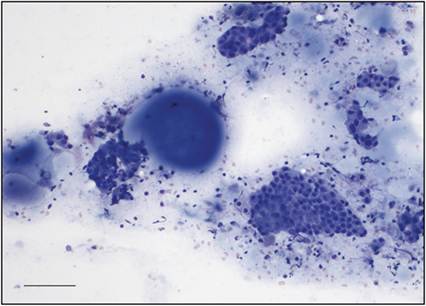
Figure 4.3 Clumps of epithelial cells and lakes of basophilic secretory material from a mammary mass in a 15-year-old, spayed female Pomeranian dog. These epithelial cells are relatively uniform in size and display few criteria of malignancy (Wright–Giemsa, scale bar = 100 μm).
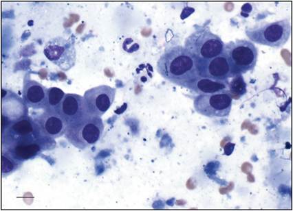
Figure 4.4 Higher magnification view of the same case as Figure 4.3, showing a clump of relatively uniform epithelial cells, a foam cell, and basophilic secretory material. Several neutrophils are also present, indicating inflammation or possibly necrosis associated with the lesion (Wright–Giemsa, scale bar = 10 μm).
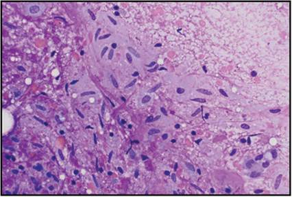
Figure 4.5 Spindloid cells admixed with granular to fibrillar pink extracellular matrix material and a few blood cells in an FNA from a mammary mass on a 7-year-old, spayed female Boxer. These cells appear most consistent with myoepithelial cells but could represent mesenchymal/stromal cells (Wright–Giemsa, 500? magnification).
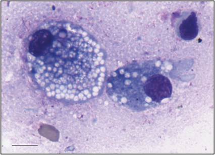
Figure 4.6 Foam cells from a mammary mass in a dog. These cells are often found in mammary gland or mammary masses and appear similar to macrophages (Wright–Giemsa, scale bar = 10 μm).
Cytology preparations of mammary lesions (both neoplastic and non-neoplastic) may also contain mammary secretions. These secretions are usually lowly cellular and contain lipid or proteinaceous secretory material, the latter of which typically appears as lakes of smooth, basophilic material (Figures 4.3, 4.4).
Foam cells may be found in mammary gland aspirates or in mammary secretions. These are round cells that resemble macrophages (Figure 4.6). They typically exhibit heavily vacuolated cytoplasm and may also contain basophilic secretory material. In humans, immunohistochemistry has shown that mammary foam cells are consistently positive for CD68 and are, therefore, of histiocytic/macrophage origin (King et al., 2002). Low numbers of macrophages and neutrophils may also be found in mammary aspirates.
Other subcutaneous glands
Please see Chapters 6 (‘Oral Cavity Cytology’) and 16 (‘Cytology of Endocrine Tissues’) for further information on salivary gland and thyroid and parathyroid gland cytology, respectively.