Syphacia muris, Syphacia obvelata and Aspiculuris tetraptera: Pinworms
Rats may serve as hosts to 3 species of pinworms: S. muris, S. obvelata, and A. tetraptera, which are generally found in the cecum and colon in infested animals. Syphacia muris commonly occurs in laboratory and wild rats and is transmissible to the laboratory mouse.
In contrast, S. obvelata is primarily a pinworm of mice that may infest rats. These parasites have a direct life cycle. Eggs are deposited in the colon or on the perianal area. The eggs embryonate and become infectious within a few hours. Rats may become infested by direct ingestion of embryonated eggs from the perianal region, ingestion of eggs in contaminated food and water or from fomites, or direct migration of larvae via the anus to the large intestine. Infestation is frequently subclinical, but younger animals with heavy infestations may exhibit various signs, including diarrhea, poor weight gains, impactions, rectal prolapse, and intussusceptions. Aspiculuris tetrap- tera frequently occurs in conventional rats and mice. The life cycle is also direct. Eggs are passed in the feces and, therefore, are not found in the perianal region.Diagnosis
The microscopic demonstration of the characteristic eggs on touch preparations of the anal region (using transparent adhesive tape) is a useful method for detection of Syphacia, but has little value for detection of Aspiculuris. Eggs of all 3 species can be demonstrated in fecal samples, and adults are visible as small, threadlike worms in the cecum and colon. Adults are also readily seen in tissue sections of large intestine, identifiable as nematodes with typical oxyurid lateral alae (Fig. 2.49). Occasionally, focal submucosal granulomas may be evident in sections of the large intestine on microscopic examination. Syphacia and Aspiculuris can be differentiated by morphologic features of the adults and eggs.
Trichosomoides crassicauda Infestation
Trichosomoides crassicauda nematodes occur in the urinary tract of wild rats and, rarely, in laboratory rats.
infested animals are usually clinically normal. The thread-like adult worms are found in the lumen and mucosa of the urinary bladder and renal pelvis at necropsy. In tissue sections examined microscopically, migratory-stage larvae and immature worms may be present in multiple tissues, particularly in the lungs.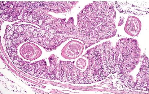
FIG. 2.49. Cecum from a rat with pinworm infestation. Note cross sections of nematodes with characteristic lateral alae.
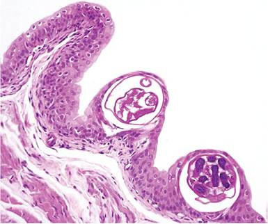
FIG. 2.50. Urinary bladder mucosa of a rat infested with Trichosomoides crassicauda (bladder threadworm). Female worms burrow into the urothelium.
Adult females reside in the epithelium of the renal pelvis and the urinary bladder (Fig. 2.50), which may elicit a chronic inflammatory response. Males are much smaller than females and live within the urinary tract lumen or within the uterus of the larger females. Typical double- operculated eggs are passed in the urine, and intracage transmission readily occurs. Urinary calculi and bladder tumors havebeen associatedwith this parasitic infestation.