Terminology and definitions
In any field of study, there are terms and expressions that are common and allow easy communication. You
Alignment steps
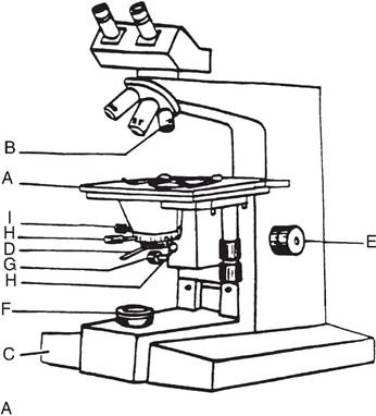
1 Carefully place a slide on the stage ( A).
2 Swing the IOx objective (B) in to position and turn on the light source (C).
3 Open the substage condenser diaphragm about 1 fourth ( D).
4 Focus on the specimen (E).
5 Close the field diaphragm (F) to a small aperture (panel B).
6 Move the substage condenser (G) up and down until the edge of aperture is in sharp focus.
7 Center the image of the field diaphragm with centering screws (H).
8 Expand the image of the field diaphragm aperture until the lighted area just fills the field of view.
9 Adjust the substage condenser (D) diaphragm for maximum resolution but do not open and close to regulate brightness (use the light source intensity).
B
Fig. 4.1. Microscope parts and use.The first step in getting the most out of your efforts is to make sure your microscope has the proper illumination.The first step is to place a prepared slide on the microscope stage (A) and carefully rotate the 10? objective lens (B) into position and then turn on the light source (C).You should then adjust the focus knob (E) to view the image.Further steps are outlined in the box highlighted in panel B. The get the best image, illumination should be centered on the specimen and the beam of light should completely fill the aperture of the objective lens. Examples of needed adjustment are detailed in Figure 4.2.
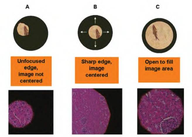
Fig. 4.2. Microscope alignment. In panel A, the field diaphragm (F) has been closed to create a small aperture; however, the image is not centered in the field of view and the edge is not in focus.
In panel B, the edge has been focused by carefully raising or lowering the substage condenser lens (G) until the image of the field of light is at its sharpest. The image has been centered by adjusting the two centering screws (H). In panel C, the field diaphragm has been opened further to expose more of the image. Letters in parenthesis refer to the microscope parts in Figure 4.1.have already been introduced to some basic physiology language in Chapter 1. As we begin to explore the structure of tissues, it is important to appreciate some of the specific language and terms that apply:
• Histology. Subspecialty of anatomy that deals with the microscopic structure of tissues.
• Tissue. A group of similar cells and intercellular materials specialized to carry out a specific activity. The four primary tissues are epithelial, muscle, nervous, and connective tissue.
• Organ. A discrete portion of the body composed of two or more tissue types dedicated to a particular function. For example, the heart is an organ of the circulatory system.
• Cytology. Subspecialty of anatomy that deals with the structure and functional differentiation of individual cells either as isolated cells or as part of a tissue.
• Pathology. Subspecialty of anatomy and physiology that deals with changes in gross anatomy, histology, or cytology associated with disease or injury.
• Necropsy. Refers to the gross and/or microscopic examination of organs, tissues, and cells after death;most often associated with determinations of the cause of death.
• Parenchyma. Refers to the functional portion of a tissue or organ. For example, in the kidney, the epithelial cells of the nephron are responsible for the formation of urine and the recovery of important nutrients filtered into the lumen of the nephron. Thus, these epithelial cells make up the critical functional structure of the kidney.
• Stroma. Refers to the support cells, that is, connective tissue, blood vessels, and nerves, that are needed for the parenchymal tissue to carry out its functions.
Although the task of learning the rudimentary histology of various tissues and organs may seem daunting at first, the job becomes easier when the information is organized into more manageable blocks.
For example, any cell or cellular product can be classified into one of four basic tissue types. Names and primary functions are outlined as follows:• Epithelium. Covering for protection, glandular activity.
• Muscle. Movement, cardiovascular function, heat production.
• Nervous. Signaling, control, integration of physiological systems.
• Connective. Support, mineral storage, protection.
We will cover the basics of each of these tissues in this chapter, but as we consider more of the physiology of various organ systems in subsequent chapters, we will often return to discuss structural attributes of cells or tissues. This is because to a very large degree, structure and function go hand in hand. In other words, the capacity of a tissue or organ to complete a specific function is directly dependent on the arrangement
Box 4.1 Tissue structure and histology: Is it ancient history?
As you have been reading the descriptions of tissues and cells and realized that many of the basic techniques have been around since the 1900s, you might be thinking, what is new in the study of tissues and cells?
First, producers and animal scientists are very interested in tools that might increase or improve efficiency of production—meat, milk, eggs, and so on. This often requires evaluations of tissues related to cell growth and development. Often histology and, increasingly, detection and quantitation of cellular proteins (receptors, signaling molecules, and cell products) are needed to evaluate the impacts of treatments and animal management to understand mechanisms. This detailed study often uses specific antibodies linked with "secondary" antibodies that contain molecules that can be induced to fluoresce or enzymatically react so that the cellular and tissue location of the protein that the specific (first antibody) recognizes can be detected and the amount of the protein quantified. Similar techniques can be used to detect and measure mRNA.
Second, domestic animals are being used as models to evaluate methods and treatments to better understand disease or injury mechanisms related to human health needs. This often requires evaluation of detailed responses of animal tissues and cells to various treatments including changes in tissue development, cell proliferation, and function. A recent review by Bartol et al. (2013) elegantly reviews the 'Tactocrine signaling" hypothesis whereby bioactive factors in mother's milk act to modify tissue development in the neonate, in this case, development of the female reproductive tract and associated expression of multiple signaling and regulator proteins.
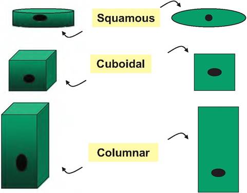
Fig. 4.3. Epithelial cell shape classifications. These stylized illustrations show three-dimensional as well as surface views for three common shapes Ofepithelial cells. The cell nucleus is indicated by the black oval. Relatively flattened, thin cells are squamous. The one cell thick row of cells that line the internal surface of capillaries or the lung alveoli are examples of squamous cells. Cuboidal cells, as the name suggests, are similar to a child's set of ABC toy blocks. The cells approximate cubes. Such cells appear as part of the lining of many ducts in glandular tissues. Columnar cells, by contrast, are more elongated and can be likened to tiny skyscrapers. These cells appear on the surface of the lining of the intestinal tract, among other places.
and organization of the cells within the tissue or organ (Bacha and Bacha, 2012) (Box 4.1).