Epithelial tissue
As illustrated in Figure 4.3, epithelial cells are classified based on the shape of the cells. In addition, the number of epithelial cells in the layer adds an additional element of classification (Fig.
4.4). A single layer of squamous, cuboidal, or columnar cells is called a simple epithelium. An alternative structure with several layers of cells is called stratified.These stylized images are oversimplified, but you should get the idea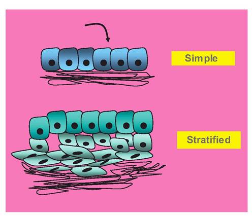
Fig. 4.4. Epithelial cells classified by number of strata. This classification is straightforward; a single layer of cells constitutes a simple layer, but when there are multiple layers of cells, this is called a stratified epithelium. The black lines represent various extracellular matrix proteins that provide anchorage and support for the layer of epithelial cells.
of how these cells are classified. One of the things that will take some practice is to distinguish epithelial cells from other cells present in tissues, for example, connective tissue cells (fibroblasts, adipocytes). One key is that the epithelial cells are often on a surface (even if the surface is internal, the lining of a duct for secretion onto the internal surfaces of body cavities). When the stratified type occurs, the shape classification is only considered for the single layer of cells on the outer surface. For example, in the stylized example given in Figure 4.4, the epithelium would be classified as stratified cuboidal epithelium.
Notice that the outer layers of epithelial cells in both examples in Figure 4.4are classified as cuboidal. Second, the dark lines underneath the cells represent the basement membrane that the epithelial cell layer rests upon. This is an unfortunate term, in that the basement membrane is not a true membrane in the usual sense, but is a complex of extracellular proteins (collagen, elastin, etc.), proteoglycans, and so forth, that serve to support and anchor the epithelial layer.
These proteins may be apparent in some histological preparations but not in others. This is depends on the fixation process used to preserve the tissue and the particular staining process (see Fig. 2.5). Since most routine processing focuses on the cellular structure, do not be alarmed if the basement membrane is not also apparent.Tissue sections prepared for the light microscope are usually made from tissues that have been preserved in formalin, dehydrated in ethanol, and ultimately infiltrated with paraffin wax, as discussed briefly in Chapter 2. These tissue blocks are then placed in a machine called a microtome that is used to cut thin slices or sections that are then mounted on a microscope slide and stained. This is how the majority of tissues have been prepared since the late 1800s. Although this is a routine process, the sections prepared can be relatively thick, sometimes more than one cell in thickness, so that you are sometimes looking at parts of multiple cells. Just like problems with other artifacts, wrinkles, tears, and so forth, thickness has to be considered as you use the microscope or consider illustrations. A further problem concerns learning to recreate three dimensions from the flat images you will be studying. Take a moment to consider the examples of what the cutting angle does to the image you see in the microscope. Imagine a real organ with twisting and turning epithelial ducts or blood vessels and the possible variations. How would this equate to the two-dimensional image of tissue on the microscope slide? For many structures, you will need to consider, has the specimen been cut longitudinally, in cross section, or perhaps at some odd tangent? All of these things impact the image that you see in the microscope. Consider the illustrations in Figure 4.5 and Figure 4.6.
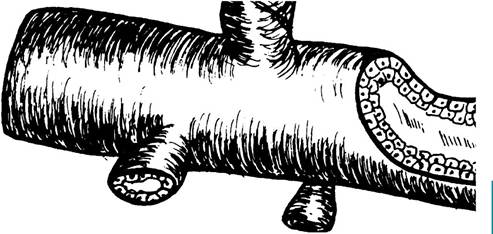
Fig. 4.5. Drawing of glandular duct. You can easily imagine that the structure illustrated in this drawing is essentially a large tube to transport secretions that empty from several other small tubes.
As the cutaway area to the right suggests, the larger tube is lined by a double layer of cells; the small cross section of one of the smaller tubes (lower left) suggests it is lined by a simple layer of epithelial cells. How would the image of the larger structure appear if it were sectioned longitudinally? What if the structure was sectioned in a perfect cross section compared with an oblique angle? What would it look like? These are some of your concerns as you examine tissue sections. You need to strive to imagine the tissue in three dimensions. 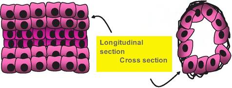
Fig. 4.6. Diagram of tubular structure. These simple drawings are an attempt to illustrate the appearance of an epithelial duct cut in either longitudinal or cross section. The cross-section profile is easier to imagine, but the longitudinal profile can easily seem like a simple mass of cells.
When you are interpreting what is seen in a single plane of section, it is important that you think about what might have been present either above or below a particular structure.
Can you now reexamine the tissue in Figure 4.7 and imagine the organization and three-dimensional structure of the tissue from the microscopic image?
Epithelial tissue characteristics
Epithelial tissue or epithelium (the plural form is epi- thelia)occurs as a sheet of cells to cover an organ surface or to line a body cavity. In other cases, epithelial tissue makes up the bulk of the cells in glandular tissues. The covering type of epithelium is abundant and widespread. These are the cells that make up the skin, the internal surfaces of the cardiovascular system, digestive tract, reproductive tract, and respiratory tract. Epithelial tissue also covers internal body cavities. The
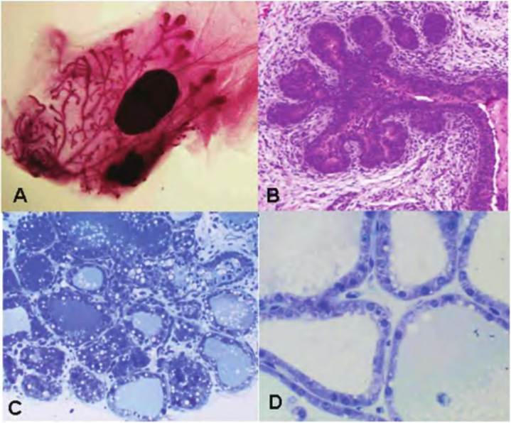
Fig. 4.7. Examples of epithelial structures. The four images shown are actual mammary ducts.
Panel A is a picture of a whole mammary gland taken from a prepubertal mouse after the gland was defatted and stained. The picture was taken with a dissecting microscope, so the ducts are intact and whole. Notice the elongated ducts with the bulbous endings (terminal end buds). Panel B is a section of mammary tissue from the mammary gland of a prepubertal Holstein heifer. Panel C is an image of mammary tissue from a pregnant heifer, and panel D is of mammary tissue taken from a lactating cow. Late in pregnancy, the mammary ducts begin to develop alveoli. The alveoli are spherical, hollow structures lined by the epithelial cells that are responsible for the synthesis and secretion of milk (this is more evident from the drawing in Fig. 4.8). The epithelial cells that line the internal surface of the alveoli are simple cuboidal. Around the outside of the alveoli, specialized myoepithelial cells form a network around the circumference of the alveolus. These cells contract in response to oxytocin released from the posterior pituitary at the time of milking. This reduces the volume of the alveolus to force accumulated milk into larger ducts and then the nipple or teat. This is called milk ejection or milk letdown. Somewhat similar structures are found in lungs, pancreas, and thyroid gland.functional cells of the accessory organs of the digestive system, that is, the liver, pancreas, and gall bladder, are mostly epithelial cells. Other glandular organs, that is, pituitary, adrenal, thyroids, salivary, and so forth, are also composed of epithelial cells. Epithelial cells form boundaries between different regions of the body. For example, the epidermis of the skin creates a protective barrier between the inside and outside of the body. The same is true for the epithelial cells that line the internal surface of the respiratory or digestive tract. Other specialized epithelial cells include reproductive cells (ova and spermatozoa), rods and cones of the retina, and the taste buds.
This explains the myriad of functions attributed to epithelial tissues: (1) protection, (2) absorption, (3) secretion, (4) excretion, (5) filtration, and (6) sensory reception.Distinctive features also contrast epithelial tissues from the other three tissue types. One of these is the degree of Cellularity of the epithelial tissues compared with that of other tissues. Specifically, epithelial tissue is composed of cells that are very tightly packed together so that usually there is a minimum of space between the cells. In fact, for the epithelial cells to successfully complete their roles as protective barriers, adjacent cells form specialized contacts. Epithelial cells acting to absorb or secrete products are described as being polarized. This is most easily visualized for glandular secretory cells. The basal region of the cell (closest to the basement membrane and capillaries) can be thought of as the manufacturing site for the cell. Products to be secreted are packaged and processed in the Golgi apparatus for subsequent secretion from the cells in the apical region of the cell (see Fig. 4.8). In other cases, the apical end of the cell (near the free surface) is acting to absorb nutrients or to move surface secretions. For example, the cells of the intestinal tract and kidney tubules have extensive microvilli. This markedly increasesthe surface area to improve function. Other epithelial cells are even more specialized with cilia—for example, cells lining the ova duct or
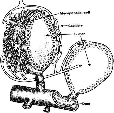
Fig. 4.8. Alveolar drawing.The figure provides a representation of the structure of mammary alveoli. As you can see, it takes experience and practice to discern the three-dimensional structure of tissues. One of the tissues for which this is very important is the kidney. Once you develop an appreciation of the structure of the kidney nephron, it will be much easier for you to understand and appreciate the filtering, reabsorption, and excretion that occurs in the urinary system.
We will now consider the features of some of the more common epithelial types.the respiratory tract that function to propel substances along their surfaces.
The epithelial cells, however, are not alone in carrying out their activities. The cells are attached to a thin supporting sheet or layer of nonliving material called the basal lamina.This layer is composed of proteins and glycoproteins that are produced by the epithelial cells. In some regions, for example, Bowman's capsule of the kidney tubules, the basal lamina is particularly thick so that it acts as a filtration barrier to prevent the movement of plasma proteins in the urinary filtrate. Underneath the basal lamina, the reticular lamina appears. This is an additional layer of more fibrous proteins, for example, collagens and elastic that link the epithelial cells with the connective tissue underneath. These two layers or lamina (basal + reticular) are collectively called the basement membrane. It defines the boundary between the epithelium and the connective tissue or stromal. Interestingly, although there are nerve fibers, that is, sensory nerves that penetrate the epithelium of the skin or intestinal tract, the epithelium is avascular. It does not contain blood vessels. These appear in the loose, more open spaces of the connective tissue. Thismeans that both the nutrients that supply the epithelial cells and waste products depend on diffusion to pass between the
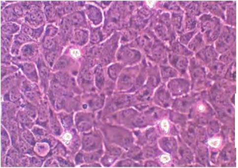
Fig. 4.9. Cultured epithelial cells. These mammary cells have formed a monolayer that is one cell thick. This is similar to the sheet of simple squamous epithelial cells that would line the surfaces of organs or surfaces of internal body cavities.
tightly packed epithelial cells and the capillaries underneath. This likely explains why it is rare to find epithelial tissue that contains more than a few strata of living cells. A final property is the capacity of epithelial cells for rapid growth and regeneration. For example, notice how quickly skin abrasions heal. But this may have a downside when you consider that most cancers are carcinomas, that is, derived from epithelial cells.
Simple epithelium
As you will likely experience in a laboratory setting with a microscope and a set of slides or in multimedia presentations, tissue samples contain multiple cell types and, in the case of epithelial tissues, often more than one type of epithelial cell. This means that while the focus of a particular specimen maybe on a specific cell or tissue, it does not mean this cell or tissue type is exclusive. Our first example (Fig. 4.9) shows epithelial cells growing on the surface of a cell culture dish. These cells have proliferated and arranged themselves into a pavement of cells one cell layer thick. If you imagined these cells growing on a flexible sheet that could be rolled into a tube, you would have a simple recreation of a capillary. Regardless, in this view, you are looking directly down onto the surface of the cells. Each cell looks something like a fried egg, with the nucleus the yolk of the egg. The cells are flattened and closely packed together. They would be classified as simple squamous. As another way to visualize simple squamous cells, imagine the flattened floor tiles in a kitchen as single squamous epithelial cells all linked together to make the floor. The grout between the tiles would represent the membrane junction complexes
that anchor epithelial cells together and create functional barriers between tissue compartments, that is, the surface and the subflooring underneath. It is typical to find simple squamous epithelium in areas where absorption and filtration occur and a thin barrier are desirable, for example, capillaries or lining of alveoli of the lungs. Can you rationalize why simple squamous cells would be a poor choice for the surface of the body?
The image shown in Figure 4.10 is from the kidney and is mostly parenchymal tissue. It shows a series of cross-sectioned tubules from several nephrons. You should remember that epithelial cells are often found on free surfaces, even though some of the surfaces may be very minute internal surfaces, that is, the inside of small vessels or tubules. Some of these tubules are lined by a single layer (simple) of squamous epithelial cells, but others are lined by a single layer of cuboidal epithelial cells. Can you find examples of each? Perhaps a hint is in order. For many of the squamous epithelial cells, the cytoplasm is very thin so that the most prominent feature of the cells is the nucleus, which often seems to protrude into the Iumenal space of the tubule. Can you pick some of these out of the image?
Figure 4.11 shows a portion of this tissue taken at a higher magnification, l,000?. This is accomplished by the use of the 100? objective lens of the microscope and the 10? eyepiece. This means that the magnification reaching your eye is 1,000-fold. The camera used to take the photographs also utilizes a 10? lens mounted in position where the eyepieces would nor
mally be located. There can also be additional magnification associated with printing or viewing, but this does not really increase true resolution. At this magnification, the difference in the cellular appearance of squamous and cuboidal epithelial cells should be apparent in the lower portion of the image. Many of
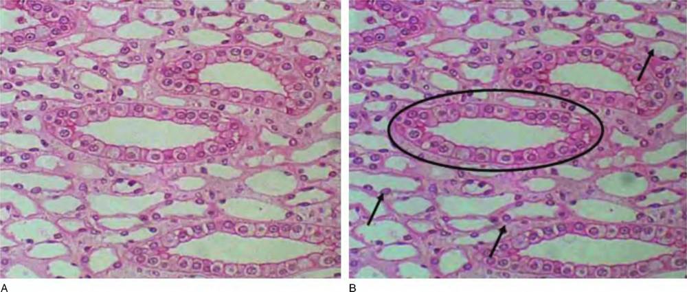
Fig. 4.10. Kidney tissue. Multiple kidney tubules are cut in cross section. Some are thin-walled regions of the loop of Henle, lined by simple squamous epithelial cells; others are sections of capillaries (arrows), also lined by simple squamous epithelial cells (called endothelial cells). There are also a smaller number of cross sections through a portion of the nephron called the collecting duct. These are lined by simple cuboidal epithelial cells (circle).
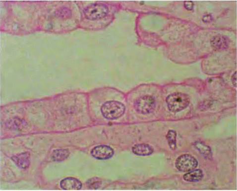
Fig. 4.11. High-power image of kidney tissue. Figure 4.12 is taken with a 20? objective and is an image of a tangential section through a blood vessel, specifically a vein. You can see clusters of red blood cells in the Iumenal space of the vessel and you can also distinguish a layer of simple squamous epithelial cells that line the internal surface of the vessel. The center of this section shows the lumen of a vein that has been cut at a tangent. Several red blood cells are clustered near the upper center of the vessel lumen. The cells that line the side of the lumen are endothelial cells.
the cuboidal cells have distinct pink staining around their borders, and the nucleus when present is generally oval-shaped and positioned in the center of the cells. Three of these cells are present near the center of the image. For the low squamous cells surrounding the lumen of a smaller duct (lower portion of the slide), the cells have only a thin rim of cytoplasm, but the nuclei are prominent and seem to protrude into the lumen of the duct.
Figure 4.12 is taken with a 20? objective and is an image of a tangential section of a blood vessel, specifically a vein. You can see clusters of red blood cells (RBCs) in the Iumenal space of the vessel. The cells that line the side of the lumen are endothelial cells. Notice the difference in the staining compared with Figure 4.10. This means you need to learn structures not based on color but on morphological characteristics.
Figure 4.13 shows a similar section through an artery at higher magnification. The box (yellow) in the figure indicates a portion of the tunica intima or interna, which is composed of simple squamous epithelial
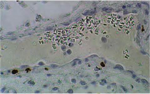
Fig. 4.12. Section through blood vessel.
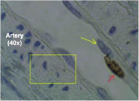
Fig. 4.13. Section of artery wall.
cells (also called endothelial cells) and the layer just under these cells, the tunica media, which has smooth muscle cells in arteries. The yellow arrow points to the nucleus of an endothelial cell and the red arrow to another endothelial cell's nucleus, which was synthesizing DNA at the time the sample was taken. The brown stain is due to the attachment of an antibody that is specific for presence of bromodeoxyuridine (BrdU) an analog of thymidine that is used to measure DNA synthesis. You should recall the relevance of these analogs in the study of cell proliferation from your earlier reading.
Before we leave our discussion of simple epithelium, as you have likely gathered from the figure descriptions, some simple squamous epithelia have specialized names. The term endothelium (meaning inner covering) is used to describe the lubricating cell covering for all vessels of the cardiovascular system including the lymphatic vessels and the internal surfaces of the chambers of the heart. Capillaries specifically are made of endothelium. This structure promotes rapid, easy movement of nutrients to the surrounding cells as the corresponding uptake of waste products. Similar simple squamous cells also make up the mesothelium (middle covering), the epithelium that makes the serous membranes of the body. These are the coverings of the internal organs and body cavities that are well lubricated to allow organs toslide past one another.
Figure 4.14 is an image from a tissue sample taken from a section of the small intestine. A portion of a villus is shown with a layer of simple columnar epithelial cells covering the outer portion. The nuclei
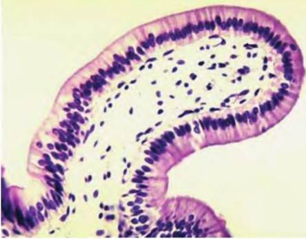
Fig. 4.14. Simple columnar epithelial cells from the intestine. This tissue section is longitudinally cut through a villus in the intestine. The epithelial cells appear as a uniform row of cells that cover the surface. Notice the dark blue-purple nuclei, most of which appear lined up in the basal region of individual epithelial cells.
appear mostly in a row in the lower third of the cell. Notice the cells are tall and narrow.
Although not apparent at lower magnification, the apical ends of the cells have many microvilli, which add capacity for absorption. This is also called the brush border. If you look closely, you should notice that the outer rim of the cells looks as if the cells have been slightly colored. This is because the microvilli clump slightly and trap proteins when the tissue is preserved. These associated proteins and carbohydrates are called the glycocalyx. The accumulated material and closely aligned microvilli allow staining and explain the darker rim.
Can you distinguish individual epithelial cells? Columnar cells are usually associated with absorption and secretion and are found lining the intestinal tract from the stomach to the rectum. This epithelium has two modifications that greatly aid its functioning. The first is the presence of the microvilli that markedly increase absorptive surface, and the second is the presence of goblet cells. These unicellular glands produce mucus that is secreted on the epithelial surface. This increases lubrication and provides protection. These specialized secretory cells also appear in the respiratory and reproductive tracts. Although the images shown in Figure 4.14 and Figure 4.15 are excellent, representative examples of the features of intestinal tract epithelium, it is important to appreciate that not all histological sections are of such quality. Also, as indicated previously, the plane of section can make it difficult to interpret a given tissue section.
The image in Figure 4.16 is also a section through intestine. It is still possible to distinguish the presence of villi and the appearance of the epithelium, but can you detect some of the problems? First, the image is a bit out of focus, and second, it is a bit too thick. This makes it difficult to distinguish individual cells. The villi have become pushed into one another during processing, so it takes some effort to distinguish individual structures. Finally, to the upper right and far right of the section, there are some tears that have altered the orientation of the tissue.
Many other artifacts also can occur. The point is that section preparation is sometimes as much an art as a science, so patience is needed as you study even professionally prepared slides. Regardless, several villi have been sectioned roughly along their longitudinal axis. This simply illustrates what you can and will see when you examine actual slides. Since you know what you are looking for in columnar epithelial cells from Figure 4.14 and Figure 4.15, you should still be able to distinguish several villi covered by a layer of columnar epithelial cells.
Figure 4.17 illustrates a similar section of the duodenum, but the sample is from a calf and the animal was injected with BrdU2 hours before the tissue was collected. Remember, this is the analog of DNA that gets incorporated into cells that are in the S-phase of the cell cycle. In this section, many of the villi are cut at a tangent to the longitudinal axis, but you can see that there are many brown-stained nuclei (indicating the cells were synthesizing DNA, i.e., presence of BrdU) in the lower regions of the villi. It is well known that the cells that populate the villus proliferate in lower crypts and are lost from the upper region of the villi as they age. To maximize the opportunity to detect labeled cell nuclei but to also be able to distinguish basic tissue structure, the sample was only briefly counterstained in hematotoxylin but without eosin. This gives the pale blue staining to the cells, but
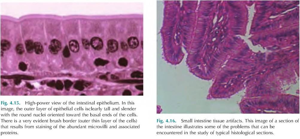
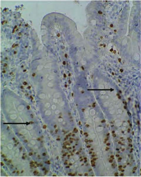
Fig. 4.17. Section of duodenum from BrdU-injected calf. This section of intestinal tissue is processed to show the presence of BrdU-IabeIed cell nuclei. Several villi are closely aligned and some are cut at a tangent, but it is apparent that the number of BrdU- Iabeled cells (brown-stained nuclei and arrows) is markedly higher in the crypts of the villi. The pale globules indicate the presence of goblet cells. Image is courtesy of Dr. Anthony Capuco, USDA, Beltsville, MD.
it is less distinct than in the H&E-stained sections of intestinal tissue (Fig. 4.14 and Fig. 4.15).
Subsequent figures,Figure 4.18and Figure 4.19, give examples of simple columnar epithelial cells from the reproductive tract. The complex folding of the internal surface is evident (Fig. 4.18), as is the regular arrangement of epithelial and goblet cells (Fig. 4.19).
Figure 4.18 shows a tissue section from the anterior (fornix) vagina of a cow taken during the follicular phase of the estrus cycle. Notice the epithelium is on the internal surface, and as the higher magnification (40? objective) image (Fig. 4.19) shows, the epithelium is also a simple columnar epithelium.
Stratified epithelium
To this point, we have considered examples of simple epitheliawith squamous, cuboidal, or columnar cells. Now let us consider stratified epithelium types. As you should surmise, the stratified types are better able to withstand physical trauma and wear and tear than
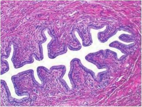
Fig. 4.18. Tissue from the anterior bovine vagina. This section illustrates the general structure of the internal lining of a region of the bovine reproductive tract. The surface is thrown into folds and is covered by simple columnar epithelial cells. Notice the red- stained, dense connective tissue surrounding the epithelium.
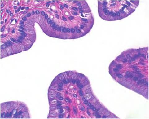
Fig. 4.19. Simple columnar epithelial cells. This section is a higher-power (40? objective) image of the epithelium shown in Figure 4.1 8. Note the layer of closely aligned epithelial cell nuclei in Figure 4.20, which shows a low-power image of sectioned cornea.
simple epithelial types but are much less efficient at absorption. This means these cells are also poorly adapted for secretion. When secretions are needed along a stratified epithelial surface, this is usually accomplished by the presence of exocrine glands that are located inferior to the epithelial surface. Ducts that radiate from the glandular cells to the surfaces provide most needed secretions. However, most of the lubrication for these internal epithelial surfaces is provided by goblet cells. As mucus accumulates in the cells, they eventually rupture to release their contents.
Remember that with stratified epithelia, the classification depends on the shape of the epithelial cells on the outer surface, adjacent to the lumen or free surface. The first image in this series (Fig. 4.20) is a section through the cornea taken at very low magnification (4?) objective. The outside of the cornea is covered by a stratified squamous epithelium and the inside by a simple squamous epithelium. The bulk of the corneal structure (arrow) consists of collagen fibers arranged in lamella that are parallel to each other as well as scattered fibroblasts.
The outer stratified squamous epithelium of the cornea is about five cells thick (Fig. 4.21). The basal
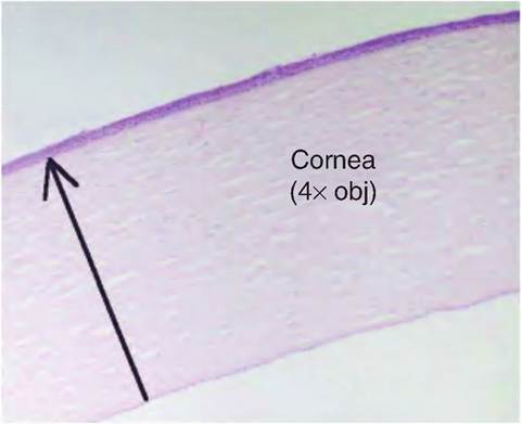
Fig. 4.20. Low-power image of sectioned cornea. At this magnification, no cellular detail is visible, but it is apparent the outer cell layer is thicker than on the inside (stratified vs. simple epithelial layers).
cells appear as cubes or polyhedrons, but the cells are progressively flattened as they migrate to the surface. Since the outermost cell layer is flatted, the classification is stratified squamous. Can you detect any of the fibroblast nuclei in the underlying substantia propria (the name given to the bulk of the corneal tissue)? Figure 4.22 shows the epithelial layer on the inner surface also at higher magnification. The cells are in a single layer and they are highly flattened, so it is an example of simple squamous epithelium. Other areas where this epithelial type would appear ison the internal surface of the lung alveoli. What better way to promote rapid diffusion of gases than with a single layer of thin epithelial cells?
Figure 4.23 provides another very common example of stratified squamous epithelium. The section is from an internal body opening that is moist but requires protection. Examples for this type of epithelium would include the lip, mouth, posterior vagina, and anus. The bracketed area indicates the epithelial portion of the tissue; the lower portion of the slide is the connective tissue or stroma. Notice the multiple layers of cells but the fact that the outermost layer of cells are flattened (arrows), therefore the stratified squamous classification. In contrast to areas that are moist, the skin also needs the protection provided by multiple layers of cells, but excess loss of moisture can be a problem for many animals. Figure 4.24 shows the beginnings of the keratinization process. The number of cell layers and classification is similar except that there is now a layer of keratin (a cellular protein) and a layer of progressively dying cells. This keratin layer provides protection. As a specific example, the keratin that is produced in the teat opening of lactating cows is a very important protection against mastitis. In experiments in which the keratin has been artificially
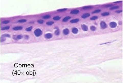
Fig. 4.21. Stratified squamous epithelium cornea. This higher- magnification view of the outside of the cornea shows several layers of epithelial cells. The outermost layers are higher flatted. Within the body of the cornea there is the faintly stained nucleus of a fibroblast (right).
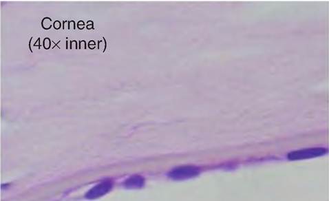
Fig. 4.22. S imple squamous epithelium cornea. At higher magnification, only a single layer of highly flattened epithelial cells is apparent on the internal corneal surface. As in the previous figure, there is also a faintly stained fibroblast nucleus within the lamellae of the cornea.
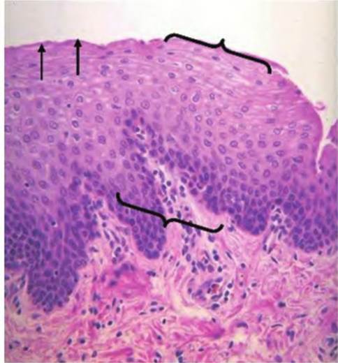
Fig. 4.23. Nonkeratinized stratified squamous epithelium. This tissue sample from just inside the bovine oral cavity shows the hallmarks of stratification, that is, multiple layers of epithelial cells (bracket area). The outermost layers of visible cells are highly flattened (arrows), thus the squamous classification. The lower area of tissue is connective tissue.
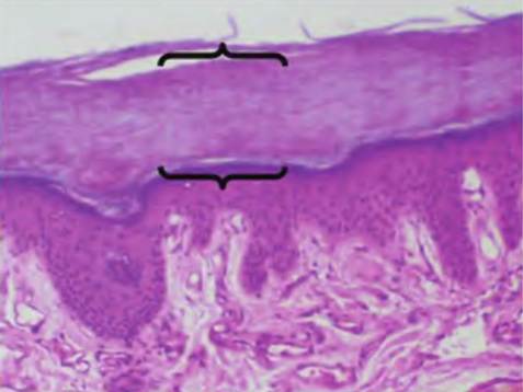
Fig. 4.25. Low-power section of skin. This low-power image shows a section of skin from a region with high friction and pressure. The outermost layer called the stratum corneum (brackets) can account for 75% of the total epithelial thickness. It is composed of keratin and thickened plasma membranes from multiple layers of dead cells.
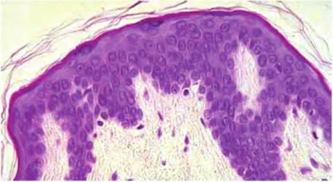
Fig. 4.24. Keratinized stratified squamous epithelium. This tissue sample from just inside the bovine reproductive tract shows the beginnings of keratinization. The outer visible layer of cells is highly flattened and more darkly stained. There are also strands of keratin fibers near the surface of the tissue.
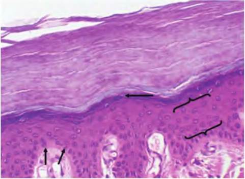
Fig. 4.26. High-power section of skin.In this higher magnification, you can see some of the morphological characteristics that distinguish other strata in the epithelium.For example, the dark- stained boundary (upper arrow) at the lower edge of the stratum corneum is called the stratum granulosum because of the presence of the keratohyaline granules. The bulk of the cells are in the stratum spinosum (brackets) and are bounded by the stratum basale (lower arrows), which appear as lighter, staining cells occurring just before the connective tissue in the dermis.
removed, incidence of mastitis is markedly increased. The next image Figure 4.25 shows a more extreme example of the protection that is provided by keratinization in the skin. Here, dead and dying cells form a very distinct outer layer that markedly increases protection against abrasion. The layer is especially increased in skin areas subjected to pressure. Figure 4.26 shows some of the cellular features of skin at higher magnification. Here you can begin to see staining and morphological characteristics that allow the epithelial cells in varying strata within the epithelium to be distinguished. These will be described in more detail in our discussion of the integumentary system.
Other types of stratified epithelium occur on the internal surfaces of some of the larger tubular structures in the body, that is, trachea, reproductive tract, and bladder. These will be considered in subsequent slides. Stratified cuboidal epithelium is usually associated
with various exocrine glands in which secretions made by the secretory cells of the gland must be transported through ducts to be emptied. The cells, which compose the walls of the ducts, generally do not produce secretions themselves but provide a passageway for products to the site of secretion. Exceptions include the ductal cells of the salivary gland and pancreas, which can act to modify secretions produced by the acinar cells. Figure 4.27 illustrates a cross section through a duct leading from a sweat gland. Note the roughly double layer of epithelial cells. The tissue surrounding the duct is mostly collagen fibrils and other extracellular matrix materials, a few fibroblasts, and blood vessel cells.
The cells in Figure 4.28 illustrate a type of epithelium found in the trachea and areas of the reproductive tract. These are pseudostratified because although the cells appear to be residing in multiple layers, in reality, each of the epithelial cells is anchored to the basement membrane. This may only be by a thin projection of cytoplasm, but since all the cells are attached,the layer only appears to be stratified. The sample is from the oviduct of a cow. Clusters of cilia are evident as tufts protruding from the apical ends of the cells.
The drawing provided in Figure 4.29 illustrates the arrangement Ofpseudostratified epithelial cells. Again, the nuclei appear at various layers, but all are attached to the basement membrane. It is also usual for this epithelial type to have goblet cells and cilia. Another example of pseudostratified columnar epithelium is
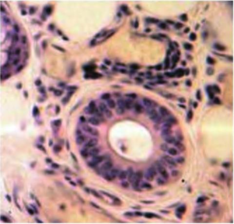
Fig. 4.27. Duct cross section. A portion of tissue from a sebaceous gland is shown. The cross-sectioned duct shows an example of stratified cuboidal epithelium. It is typical of the structure of various ducts of exocrine glands.
shownin Figure 4.30. This sample is a cross section through the epidymis of a bull. In this tissue, there is a more complex surface specialization, called stereocilia. These structures, similar to but more elaborate than simple cilia, are evident as the elongated spikes that protrude into the Iumenal space of the tubule. The center of the lumen is also filled with stored spermatozoa, a highly specialized epithelial cell.
A final epithelia type (Fig. 4.31) we will consider is transitional. This type appears in the lining of the bladder and is unique because its appearance changes
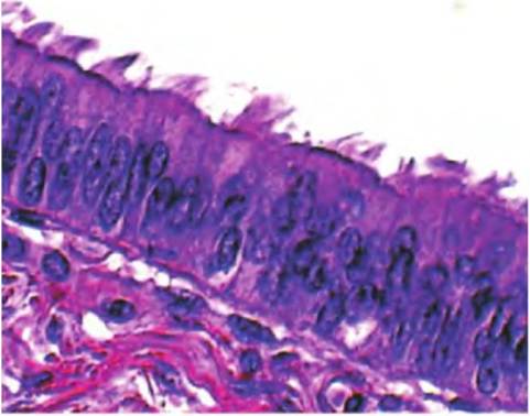
Fig. 4.28. Pseudostratified columnar epithelium. This image is from a section of the oviduct of a cow. Only one side of the oviduct is shown with the surface epithelial projecting into the lumen. The nuclei stained in dark blue-purple appear to be aligned in multiple layers. However, all of the individual cells are actually anchored in the region of the basement membrane.For this reason, the cells are classified as pseudostratified (false stratification). The shape of the cells is columnar.ln addition, these cells exhibit a surface specialization, the presence of cilia. Here they were clumped into tufts when the tissue sample was processed.
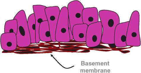
Fig. 4.29. Drawing of a pseudostratified epithelium.
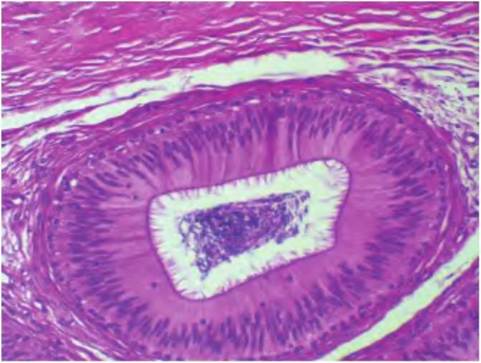
Fig. 4.30. Pseudostratified epithelium bovine epidymis. These epithelia have stereocilia that protrude from the apical cell surfaces into the Iumenal space. The movement of these stereocilia maintains the flow of maturing sperm cells in the reproductive tract. Compacted sperm cells appear as the dark cluster in the center of the Iumenal space.
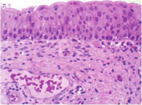
Fig. 4.31. Transitional epithelium bladder. The appearance of this epithelial layer changes related to the degree of stretch. As the bladder or other areas of the urinary tract expand, the apparent number of epithelial cell layers decreases until the pressure is relieved.
as the bladder expands and contracts. When relatively empty, the epithelium is similar in appearance to nonkeratinized stratified squamous. As the bladder fills, the expansion reduces the number of apparent cell layers. In the nondistended state, the rounded surfaces of the epithelial cells seem to protrude into the Iumenal space.Notice the cross section of the vein and artery just below the epithelium. The nuclei of the larger circumference of the vein (upper) also provide an excellent example of simple squamous epithelium.