The inner ear
The inner ear (Fig. 5.19) consists of:
The bony labyrinth, which lies within the petrous temporal bone. It is divided into three areas - the bony cochlea, bony vestibule and bony semicircular canals - each of which contains a corresponding part of (he membranous labyrinth.
It contains perilymph which flows around the membranous labyrinth. T'he bony labyrinth is linked to the middle ear bv Iwo membranes:- Round ∖vinιknv- between the bony cochlea and the centre of the tympanic cavity
- Oval window - between the bony vestibule and the dorsal part of the tympanic cavity: the stapes is in contact with (his membrane.
The membranous labyrinth is a system of interconnecting tubes filled with a fluid known as endolymph. Inside (he structure are sensory receptor cells adapted to respond to sound and movement. The shape of the membranous labyrinth corresponds to the bony labyrinth but does not completely Iill it. There are three parts: - Membranouscochlea - this has a similar shape to that of a snail’s shell. It forms a blind-ending ventral spiral and is filled with endolymph. Within the cochlea are a group of sensory receptor cells forming the spiral organ of Corti, whose function is to detect sound. Projectingfromcach receptor cell is a sensory hair and al the base is a nerve fibre which is part of (he cochlear branch of the VeslibuIiKcx hIear nerve IVH I.
Perception of sound Sound waves, picked up by the pinna and transmitted across the ear by the tympanic membrane and auditory ossicles to the oval window, set up vibrations or ripples first in the perilymph and then the endolymph. These ripples move the sensory hairs of the receptor cells in the organ of Corti, and this sends nerve impulses along their associated nerve fibres.The impulses are carried by the cochlear branch of the vestibulocochlear nerve (VIII) to the auditory cortex of the cerebral hemispheres, where they are interpreted as sound.
- Membranous vestibule - this is made of Iwo sac- like structures, the Uiricle IΓig. 5.20) and saccule. which connect with the cochlea and the semicircular canals and are tilled with endolymph. Both these structures contain areas of sensory hair cells known as maculae, surrounded by jelly-like material containing calcium carbonate particles or otoliths. The function of these structures is to maintain balance when the animal is standing still.
Stobc balance. When an animal is Stondrng stιW. the head still makes minute movements. The pull of gravity moves the jelly-like material within the maculae of the utricle and saccule.This moves the sensory hairs, which transmit nerve impulses to the brain via the vestibular branch of the vestibulocochlear nerve (VIII).The brain interprets this information in terms of the position of the body in relation to the space around it
- Membnmous semicircular canals - these are three canals, filled with endolymph, each of which describes ¾ of a circle. The plane of each circle is approximately at right angles to the other two - in this way. the three dimensions of movement are monitored. Each canal is connected to the utricle by a swollen area known as an ampulla. Inside each ampulla is a cone-shaped projection or crista containing sensory hair cells embedded in a jelly-like cupula. The function of these Slriiclures is Io maintain balance during movement.
Dynamic balance. As the animal moves, the endolymph in the semicircular canals moves and stimulates the cristae within the ampullae. Nerve impulses are transmitted by the hair cells to the brain via the vestibular branch of the vestibulocochlear nerve (VlII).Within the brain, they are passed to the cerebellum where the information is coordinated.
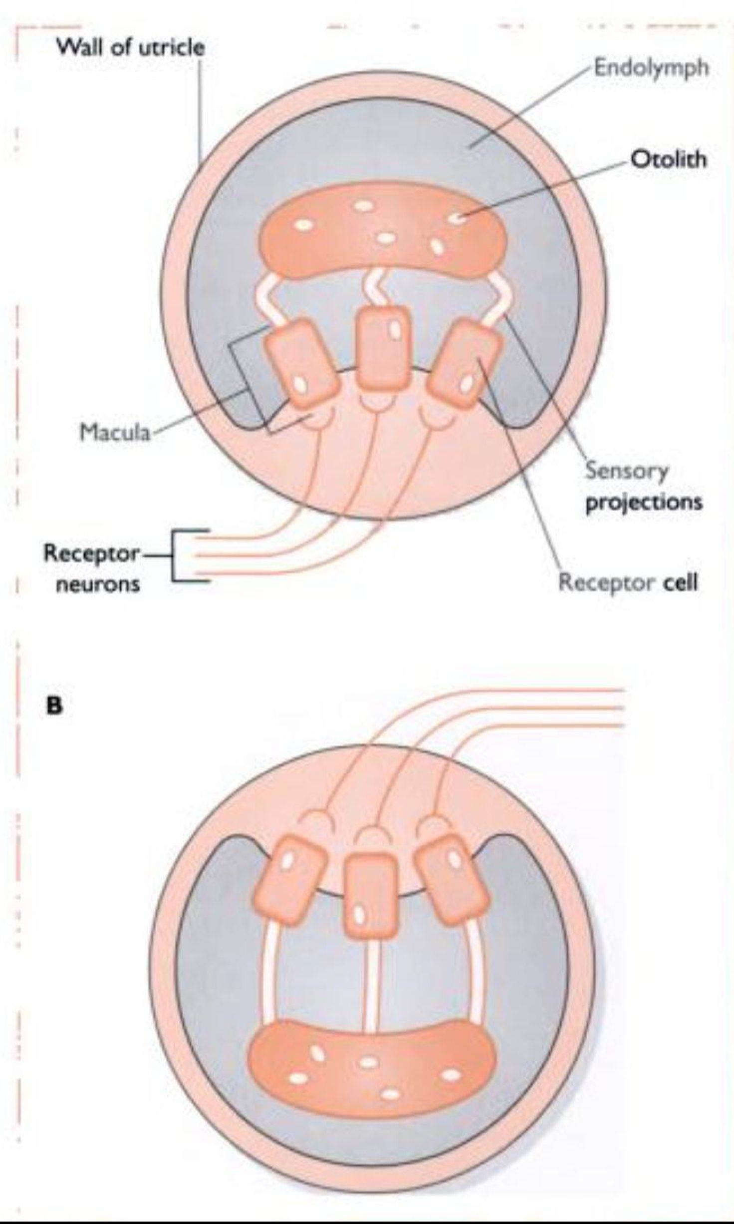
Fig. 5.20 Simp ficd section through a utnc∣e A With the head upnght. there in response to low levels of blood oxygen. It stimulates the bone marrow to produce erythrocytes or red blood cells.