The ribs and sternum
The ribs form the walls of the bony thoracic cage that protects the organs of the chest. There are 13 pairs of ribs in the dog and cat. which articulate with the thoracic vertebrae (Fig.
3.12). ∕∖ rib is a flat bone consisting of compact bone on the outside packed withThe synovial joint between the atlas (C I) and the occipital condyles of the skull allows nodding movements of the head, and the synovial joint between the atlas and axis (C I -C2) allows a pivotal movement so that the head can turn in all directions.
During parturition, under the influence of the hormone relaxin, the sacroiliac ligament relaxes and softens so that the pelvis can stretch, enabling the fetuses to pass out through the birth canal.

cancellous bone on the inside. Each rib has a bom dorsal part and a cartilaginous ventral part - th< costal cartilage. 'Γhe most dorsal part of the bony ril has two projections: the head, which articulates will the costal fovea of the vertebra, and the tubercle or neck which articulates with the transverse Jbvea of th∣ appropriate thoracic vertebra.
The costal cartilage articulates with the sternum, either directly or indirectly. The Iirst eight pairs of ribs attach directly to the sternum and are called the Ster- nal ribs. The ribs from pairs 9-12 are called asternal or false' ribs, and they attach via their costal cartilages to the adjacent rib. forming the costal arch. The last ribs (pair 1 1) have no attachment at their cartilaginous
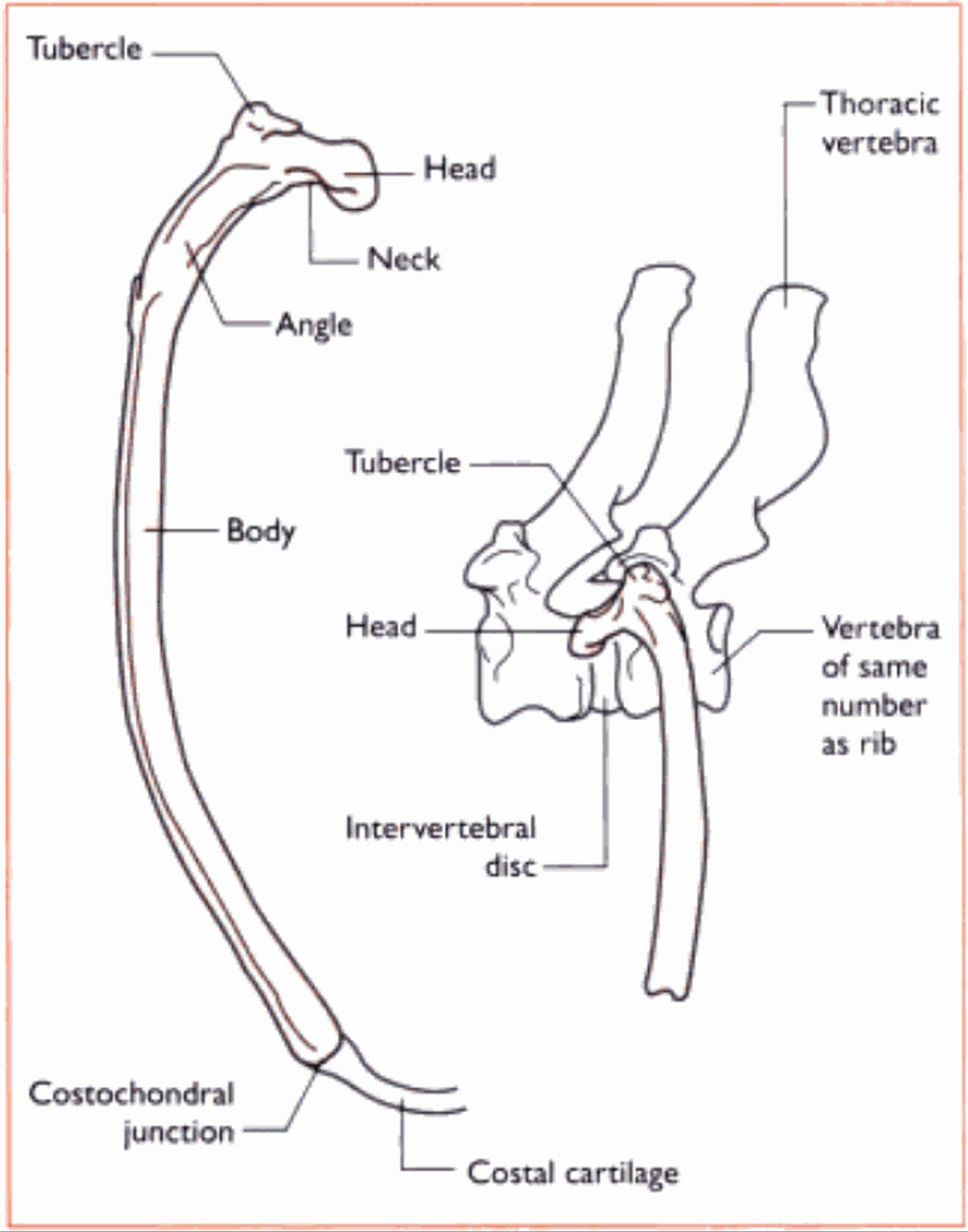
Fig. 3.12 Structure of the canine nb A Caudal view. B Lateral ⅛∣ew showing artκu∣at∣o∩ with a thoracic vertebra. (Repnnted from C∣∣n∣cal Anatomy and Physiology for Veterinary Technicians.
T Colville and JM Bassett, p 112. Copyright 2002. with permission from E∣sev∣er Science.)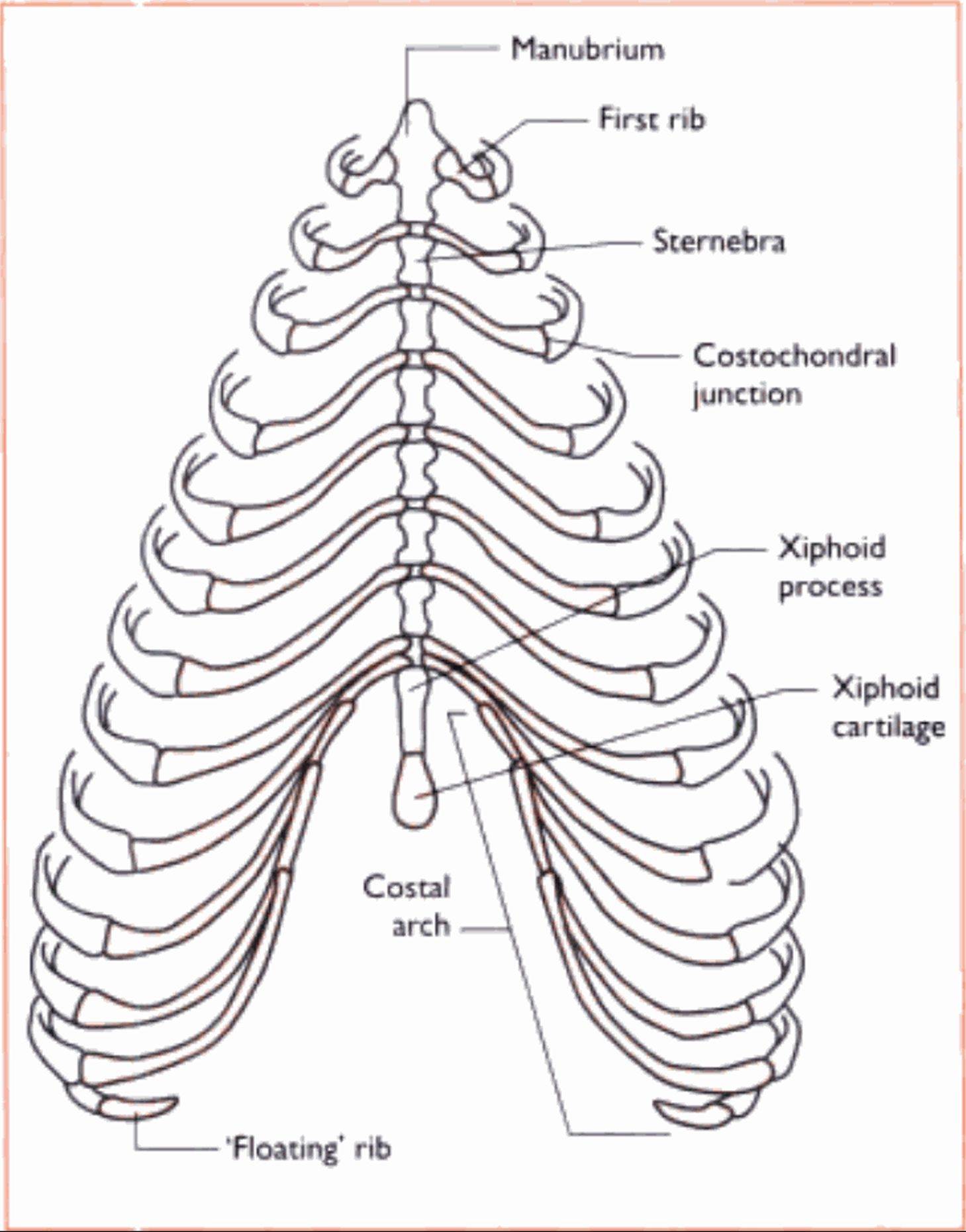
Fig. 3.13 The «ib cage of the dog. showing sternal, asternal and ’floating r∙bs. (Reprinted from Q∣n∣ca∣ Anatomy and Physiology for VeterinaryTcchnicians.! Cotvillc a∩d JM Bassett, p I IZCopynght 2002. with permission from Flsevier Science.)
ends, which lie free in the abdominal muscle - this pair are called the 'floating' ribs. T he space between each successive rib is called the intercostal space and is filled by the intercostal must les of the trunk I Fig. J. I 3).
T he sternum forms the lltκιr of the thoracic cage (Fig. 3.1 5). and is composed of eight bones, the Sierne- hrae. and the inIersternebral cartilages. The most cranial Sternebra is the manubrium, which projects in front of the first pair of ribs and forms part of the cranial thoracic inlet. Slernebnie 2-7 are short cylin-
■ drical bones. The last Sternebra is longer and dorsoven- Irally Ilallened and is called the.Viplioid process. Attached to the xiphoid process and projecting caudally is a flap of cartilage called the xiphoid cartilage. The Iinea alba attaches to this, between each Sternebra arc cartilaginous discs called the Intersternebral cartilages.