Thymus
Normal thymus is not usually accessible for aspiration. Impression smears contain large numbers of small, well-differentiated lymphocytes. Thymic epithelium can be observed. Other features include Hassall’s corpuscles, which are tight clusters of epithelioid macrophages with cell junctions, and moderate numbers of mast cells.
Thymic enlargements may be the result of some different neoplastic and non-neoplastic conditions. Thymic hemorrhage is a rare non-neoplastic disease condition that may lead to severe transitory enlargement of thymus due to blood accumulation in the context of thymus. FNAs allow collection of a highly hemodiluted sample with an admixed population of mature T lymphocytes and fewer blasts showing different immunophenotypes via flow cytometry. Among neoplastic diseases thymic (mediastinal) lymphoma and thymoma are the most frequent and should be differentiated also in light of different therapeutic approaches (chemotherapy vs. surgery) and prognosis.
Thymic lymphomas are frequent in young cats and are often related to feline leukemia virus infection (Vail, 2013). The main lymphoid population is often composed of small to intermediate lymphocytes with cleaved nuclei with or without a prominent nucleolus. Occasionally, large blasts may be the prevalent lymphoid population and help to differentiate lymphoma from other lymphocytic-rich neoplasms (Figure 5.81). Paraneoplastic hypercalcemia may be seen. Prognosis with thymic lymphoma is generally poor, with a median survival time of 2–3 months in cats (Vail, 2013).
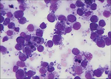
Figure 5.81 Cat, FNA from a mediastinal mass. Presence of a single population of large lymphoid blasts with prominent nucleoli consistent with high-grade thymic lymphoma. Phagocyting macrophages and mitosis are also present (Wright–Giemsa, 600? magnification).
Thymoma is also a cause of thymus enlargement. Thymic epithelial cells with some characteristics of malignancy may be observed. Cytologically, a thymoma consists of small lymphocytes with few large lymphocytes, mast cells, and occasional clusters of thymic epithelium (Figure 5.82). The ratio between lymphocytes and epithelial cells in histologic sections may be highly variable and is used to define different thymoma subtypes. Paraneoplastic conditions associated with thymoma include myasthenia gravis, hypercalcemia, megaesophagus, pemphigus foliaceous, superficial necrolytic dermatitis, exfoliative dermatitis, and pure red cell aplasia. Flow cytometric detection of double positive (CD4+CD8+) lymphocytes indicates the cells arose from a thymic mass, rather than an enlarged mediastinal lymph node. In dogs, finding a highly prevalent population of double-positive (CD4+CD8+) thymocytes is strongly suggestive of thymoma (Lana et al., 2006b). In cats, a high percentage of thymic lymphomas shows a double positive (CD4+CD8+) immunophenotype (Bernardi et al., 2020). Thus flow cytometric results do not help differentiate thymic lymphoma from thymoma. If clinically warranted, PARR is recommended to determine if the lymphocytes are neoplastic.
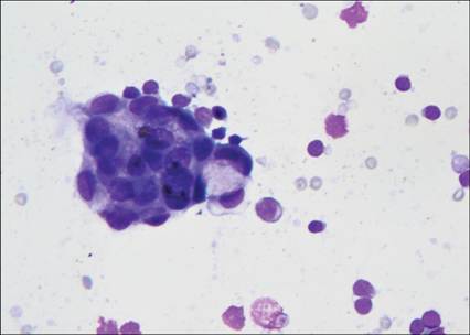
Figure 5.82 Dog, FNA from a mediastinal mass. Presence of a mixed lymphoid population with a single cluster of epithelial cells consistent with thymoma (Wright–Giemsa, 1,000? magnification).