INTRODUCTION
Human immunodeficiency virus type 1 (HIV-1) infection is characterized by a progressive loss of CD4+ T cells, and apoptosis has been proposed to be the primary mechanism involved in this process.1-2 A growing body of literature suggests that the HIV-1 transactivator protein, Tat, plays a key role in HIV-1-mediated cell death and induces apoptosis in T lymphocyte cell lines as well as in peripheral blood mononuclear cells (PBMCs).
Tat has also been shown to possess pro-survival properties. Other HIV-1 proteins have been implicated in regulating apoptosis namely Env, Vpr, and Nef.3-5 In this chapter, we will focus our attention on Tat, a small regulatory protein with pleiotropic properties that regulates apoptosis of infected and noninfected cells by a variety of possible mechanisms.HIV-1 Tat is a well-conserved protein. Although the laboratory HIV strains produce an active 86-amino acid Tat protein, most Tat proteins from primary isolates contain an additional 15- to 16- residue-long sequence at their C-terminus. Tat is encoded by two exons. The first encodes residues 1 to 72 and is classically described as a modular protein (Figure 9.1). Tat contains six regions, namely, the proline-rich N-terminus (aa 1 to aa 21), the cysteine-rich region (aa 22 to aa 37), the hydrophobic core region (aa 38 to aa 48), the basic region (aa 49 to aa 59), the glutamine-rich region (aa 60 to aa 72), and the C-terminus encoded by the second exon and with size that corresponds to the isolate. Tat displays sequence variability from isolate to isolate but contains relatively conserved residues or sequences, including a conserved tryptophane residue at position 11; seven cysteine residues at positions 22, 25, 27, 30, 31, 34, and 37 in the cysteine-rich region; the sequence 43LGISYG48 in the core region; and the sequence 49RKKRRQRRR57 in the basic region.
The functions of several regions of the protein have been well characterized.6-13 The N-terminal region 1-9 is involved in the binding of Tat to CD26 (DP IV8,11), and the region 49-57 is involved in Tat uptake. Albini et al.9 demonstrated that the synthetic peptide 24-51, which encompassed the “chemokine-like” region of Tat, induces the rapid and transient Ca++ influx in monocytes and macrophages, analogous to β-chemokines. This peptide is angiogenic in vivo.14 Boykins et al.10 showed that the fragment 21-41 is sufficient to transactivate, induce HIV replication, and trigger angiogenesis. Mutations at the positions 22 or 22/37 lead to a protein that lacks transactivation capacities and that displays a transdominant negative phenotype.15 Acetylation of Tat at positions
143
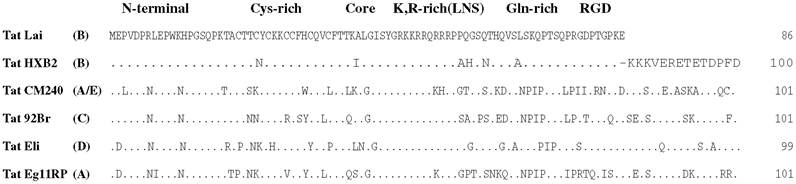
FIGURE 9.1 Sequences and modular structure of Tat proteins. HXB2 HIV-1 strain was isolated in France, CM240 in Thailand, 92Br in Brazil, Eli in the Democratic Republic of Congo, and Ug11RP HIV-1 strain in Uganda. (Adapted from Opi, S., Peloponese, J.M. Jr, Esquieu, D., Campbell, G., de Mareuil, J., Walburger, A., Solomiac, M., Gregoire, C., Bouveret, E., Yirrell, D.L., and Loret E.P., J. Biol. Chem., 2r72, 35915-35919,
2002. With permission.)
K28 and K50 is particularly important for its transactivation activity.16-18 Mutation and competition experiments, as well as ribonuclease protection assays, demonstrated that the arginine-rich motif of Tat (aa 49 to aa 57), and especially the arginine residue 52 (Figure 9.1), is essential for its high- affinity binding to transactivation response (TAR) element RNA, a 59-nucleotide stem-loop structure located at the 5' end of all transcripts.19 It is through its interaction with TAR that Tat acts as a powerful transcriptional activator.
It seems that in addition to residues present in the basic domain, the N-terminus region of Tat is also important for Tat-TAR interaction in vitro.20 In vivo, the N-terminal domain of Tat (aa 1 to aa 48) interacts directly with cyclin T1, a regulatory partner of cyclin-dependent kinase 9 (Cdk9) in the positive transcription elongation factor (P-TEFb) complex, and Tat∕cyclinT1 in the P-TEFb complex binds TAR RNA cooperatively. The assembly of the Tat/TAR/P-TEFb complex to the HIV promoter activates the Cdk9 activities, which further autophosphorylate the P-TEFb complex and hyperphosphorylate the C-terminal domain of RNA polymerase II, which leads to the formation of processive elongation complexes that synthesize full-length HIV viral mRNA.21∙22 Recent studies have shown that the nascent P-TEFb complex contains other RNA and proteins of known or unknown identity.Regarding the C-terminal domain of Tat, Poggi et al.13 reported that HIV-1 Tat and, particularly, the peptide 65-80 inhibit natural killer (NK) cell-mediated lysis of dendritic cells (DCs) and interferon (INF)-γ release by NK cells after their contact with DC. Finally, the RGD motif present in residues 78 to 80 binds the integrins α5β 1 and αvβ3, the receptors of fibronectin and vibronectin, respectively.23,24
A panoply of peptide analogues or molecules targeting specific regions of Tat as well as TAR and cyclin T1 was used as possible blocking agents in therapeutic strategies (see Hamy et al.25; Fulcher and Jans26; Holmes, Arzumanov, and Gait27; Bai et al.28; and Tikhonov et al.29 for examples).