External genitalia
The external genitalia or vulva consists of the mons pubis, clitoris, urethral opening, vestibule, hymen, labia minora, labia majora, and Bartholin's glands (Box 3.2). The functions of the external genitalia are to provide a protective cushion during sexual intercourse, to help with lubrication during sexual intercourse, to provide a protective barrier to the internal genital organs, to provide an area for the excretion of urine, and to secrete pheromones.
The mons pubis is a rounded mound of fatty tissue that lies over the pubic bone (Figure 3.2). The two labia majora are the two most lateral structures and also consist of fatty mounds. During puberty,
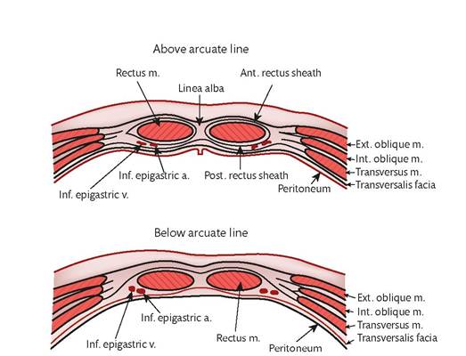
Figure 3.1 Anterior abdominal wall. a, artery; m, muscle; v, vein.
• Mons pubis
• Labia majora
• Labia minora
• Clitoris
• Clitoral glans
• Clitoral hood
• Clitoral frenulum
• Vestibule
• Urethral opening
• Skene's glands and ducts
• Bartholin's glands and ducts
• Introitus
• Hymen
• Fourchette
• Perineum
the mons and labia majora enlarge, become hair bearing, and contain sebaceous glands.
The labia minora lie just inside the labia majora and surround the opening to the vagina and the urethra (Figure 3.2). During sexual stimulation, the blood vessels of the labia minora become engorged, making them more sensitive. The clitoris lies between the two labia minora at their upper pole and is a small protrusion that corresponds to the penis in the male. It consists of the clitoral glans, the clitoral hood which is a small covering of skin, and the clitoral frenulum. The clitoris can become erect like the penis and is very sensitive to sexual stimulation, resulting in orgasm.
The vulval vestibule is the area bordered by the labia minora laterally, clitoris superiorly, and the fourchette inferiorly (Figure 3.2).
The sides of the vestibule are bordered by Hart's lines which are on the inside of the labia minora and are the transition between the vulval skin and the softer epidermis of the vestibule. The vestibule is the location for the opening of the vagina and Bartholin's glands as well as the opening of the urethra and Skene's ducts.The vaginal opening is called the introitus (Figure 3.2). This serves to function as the entrance for the penis during sexual intercourse, an exit for menstrual blood during menstruation, and an opening for delivery of a baby. The entrance to the upper vagina is bordered by the hymen which is a membranous structure. Sexual intercourse and childbirth normally disrupt the hymen, leaving remnants called carunculae myrtiformis. Bartholin's glands lie either side, inferior and posterior to the introitus, just caudal to the hymen. They produce a thick secretion that functions as a lubricant for sexual intercourse.
The urethra opens into the vestibule inferior to the clitoris and superior to the hymen (Figure 3.2). Its function is to carry urine to the outside. It is bordered by two duct openings from Skene's glands (periurethral glands). These are homologous with the prostate in the male. The function is unknown and they may be an embryological remnant of no importance. The anatomical location is very variable. It has been postulated that the Skene's glands are the site of fluid production when female ejaculation occurs.
The area between the fourchette (posterior part of the introitus) and the anus is called the perineum (Figure 3.2). This area of skin is sensitive to stimulation and may play a role in sexual arousal. The
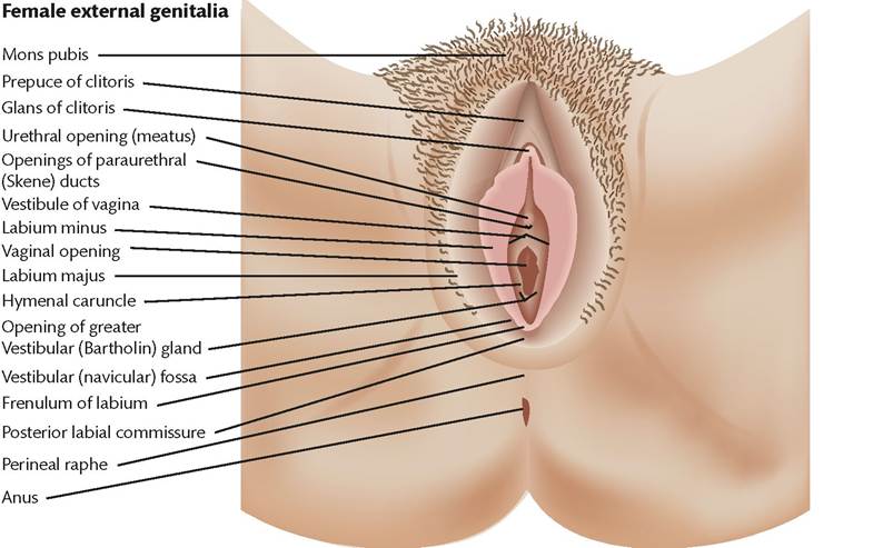
Figure 3.2 Female external genitalia.
length varies from 0.5 to 2.5 cm and can be traumatized during childbirth.
Clinical considerations
The vulva surface is a skin similar to that in other parts of the body.
For this reason, it is vulnerable to any dermatological condition that can occur in other locations such as neoplasia, infection, and dermatitis. Itching of this area is called pruritus vulvae, and pain is called vulvodynia (see Chapter XX).Bartholin’s ducts and Skene's ducts can become blocked resulting in cyst formation. If these cysts become infected, abscesses can form that might require intervention (see Chapter XX).
The labia minora vary in size immensely. Some are only a few millimetres long while others can be many centimetres. This has resulted in a fashion for cosmetic surgery to the labia minora to make them smaller. However, the term ‘labial hyperplasia’ is often incorrect and just describes normal organs that are larger than others and such surgery is usually unnecessary. lateral surface is attached to the Fallopian tube by the mesosalpinx and suspensory ligament which contain further vessels and nerve fibres.
The blood supply to the ovaries is via the ovarian artery that arises from the aorta and descends into the pelvis within the infundibulopelvic ligament (also called the suspensory ligament). The venous drainage mirrors the arterial supply. On the right side, the vein drains into the inferior vena cava. On the left side, the vein drains into the renal vein. Anastomoses occur with branches from the uterine artery within the mesosalpinx. The lymphatic drainage is via lymph vessels associated with the ovarian artery to the paraaortic nodes, in addition to iliac nodes via lymphatics following the anastomoses with the uterine artery.
Fallopian tubes
The Fallopian tubes are 10-12 cm long and pass from the superior angle of the uterus alongside the ovary. The attachment to the ovary is called the mesosalpinx and contains blood vessels and nerves that supply the ovary and the Fallopian tube.