Internal genital organs
The internal genital organs consist of the ovaries, the Fallopian tubes, the uterus, and the vagina (Figure 3.3). These all lie in close proximity between the rectum posteriorly and the lower urinary tract anteriorly.
Ovaries
The ovaries are white, almond-shaped structures that are situated behind the broad ligaments on each side of the pelvis within a depression called the ovarian fossae. The superior pole is attached to a sheet of tissue containing vessels and nerves called the infundibulopelvic ligament. The inferior pole is attached to the uterus by the ovarian ligament. The medial surface lies free within the pelvic cavity and the
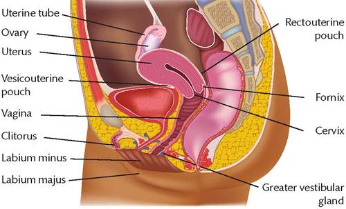
Figure 3.3 Sagittal section of internal genital organs.
There are six main parts to the Fallopian tube. These are the ostium, interstitial portion, isthmus, ampulla, infundibulum, and fimbriae. The ostium is the opening of the tube into the uterine cavity. The interstitial portion is the part that lies within the myometrium of the uterus. The isthmus is the narrowest part of the Fallopian tube that connects with the interstitial portion. The tube is less narrow at the ampulla and connects with the isthmus with the opening near the ovary called the infundibulum. This contains a number of frond-like structures called fimbriae.
The blood supply to the Fallopian tubes is via branches from the ovarian and uterine arteries. The lymphatic drainage is to the internal and external iliac nodes and para-aortic nodes.
Uterus
The uterus is divided into the cervix and corpus. The corpus is the upper part of the uterus and is lined by endometrium that sheds during each menstrual cycle. The most superior part is called the fundus. Laterally, the Fallopian tubes extend from their ostia (opening) within the uterine cavity.
The cervix is the lower part of the uterus.
The part of the cervix that projects into the vagina is called the portio vaginalis and the outside that can be seen within the vagina is the ectocervix. The cavity of the cervix (endocervix) starts from the external os within the vagina and extends to the internal os before entering the uterine cavity.The blood supply to the uterus is via the uterine artery and ovarian artery. The uterine artery divides into a superior and inferior branch after it passes over the ureter. The superior branch passes upwards and anastomoses with branches from the ovarian artery. The inferior branch is the vaginal artery. The lymphatic drainage is to the obturator, internal iliac, and external iliac nodes and via the chain associated with the ovarian artery to the para-aortic nodes.
Vagina
The vagina is a tubular structure and extends from the cervix to the introitus. The walls of the vagina come together from anterior to posterior in the upper two-thirds and from left to right in the lower third. The arterial supply is from vaginal arteries from the uterine and internal iliac artery.
The lymphatic drainage in the upper two-thirds is via the obturator, internal iliac, and external iliac nodes. Lymphatics of the lower third of the vagina drain via the vulva to the femoral nodes.
Clinical considerations
The Fallopian tubes, uterus, cervix, and upper vagina develop in utero from the Mullerian ducts on both sides. A variety of malformations can occur when the two Mullerian ducts fail to join or the system fails. They range from uterine and vaginal agenesis to a bicornuate uterus and vagina and minor abnormalities of the uterine cavity. Mullerian malformations are frequently associated with abnormalities of the renal tract. This area is covered in Chapter XX.