Management of pelvic organ prolapse
The choice of management of POP varies based on the type and severity of symptoms, combinations of the compartments involved, coexisting urinary or bowel symptoms and the risk of developing new ones, and patient factors including general health, BMI and most importantly their expectations from treatment.
In addition, the wish to retain sexual function might influence the choice of treatment.Conservative management
Pelvic floor physiotherapy/pelvic floor muscle training can help improvement of symptoms of bulge and also any coexisting urinary or faecal incontinence (26, 27). In a randomized controlled trial (RCT) on the cost-effectiveness of pelvic floor muscle training versus watchful waiting, pelvic floor muscle training resulted in a significant improvement of pelvic floor symptoms compared to watchful waiting (43% for pelvic floor muscle training vs 14% for watchful waiting) in women with stage 1 and stage 2 prolapse (28). Evidence from the multicentre POPPY Trial on supervised pelvic floor muscle training versus no intervention suggests a greater reduction in patient-reported prolapse symptoms at the end of 1 year. Though there was some reduction in the quantification of prolapse on POP- Q scoring, the difference was not statistically significant (26).
Mechanical support
Rings and pessaries of various sizes and shapes are useful in patients who do not wish, or are not medically fit, to undergo surgery. In some cases they may be used as a trial to assess the effect of pelvic organ support on the prolapse symptoms, for example, in those patients where it is unclear if a sensation of heaviness is due to prolapse or another pathology (e.g. central sensitization). Ring pessaries but not Gelhorn or shelf pessaries, can be used in women who are sexually active. The majority of the women experience resolution of bulge symptoms with nearly 50% reporting improvement in concomitant urinary symptoms.
Occult stress incontinence may get unmasked following pessary insertion (21%) resulting in dissatisfaction (29).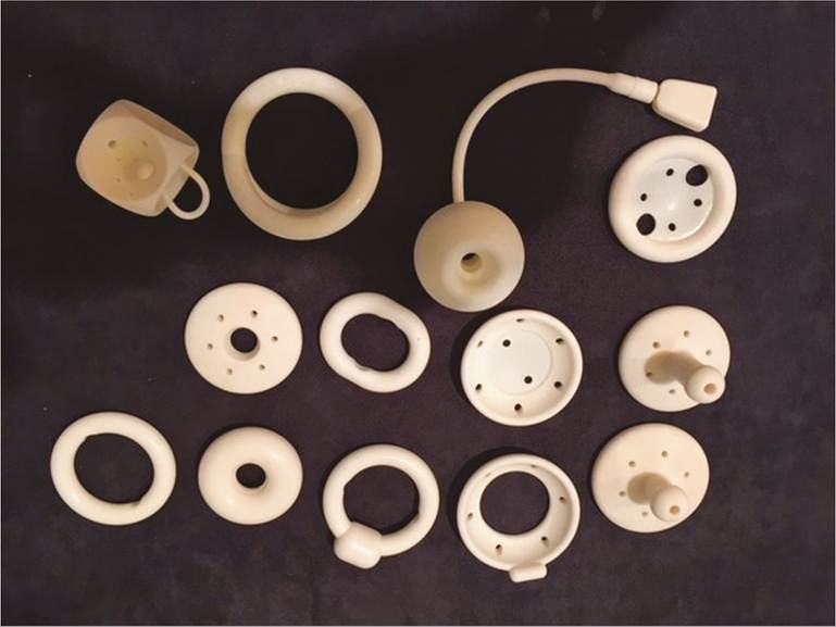
Figure 56.4 Various types of pessaries.
Shelf, Gelhorn, doughnut, and cube pessaries are space occupying and may be used in women who are not sexually active. In a randomized trial comparing pessary use to surgical management of prolapse, no difference was found in subjective outcomes at the end of 1 year of follow-up (30, 31). Patient satisfaction with a pessary after 12 months of use was found to be 59.2%. In women who manage to retain the pessaries, only 28% were found to continue using pessaries at the end of 5 years of follow-up (31, 32). The rationale for discontinuation included failure to retain the pessary, expulsion of the device while performing daily activities, discomfort, a desire for surgery, and an inability or pain during insertion/ removal the pessary. Women with enlarged genital hiatus, higher-order vaginal parity, and previous hysterectomy may not be able to retain certain types of pessaries (Figure 56.4) (32).
Women who continued to use a pessary for up to 12 months complained of vaginal discharge, infection, and, in certain cases, ulcers in the vaginal epithelium due to contact with the pessary (33). Younger women who had a previous POP surgery and recurrent prolapse had a higher rate of discontinuation of pessary treatment (34).
Minor complications were reported in 12% of the pessary users and included pain, bleeding, ulceration or excoriation, impaction, and constipation during long-term follow-up (32). Rare complications from ‘neglected'/forgotten pessaries (i.e. patient is not seen for routine follow- up and pessary change), include fistula (vesicovaginal and rectovaginal), perforation of the vault, and embedment of the pessary with a band of granulation tissue growing on top (35).
Low-dose vaginal oestrogens may alleviate some of these undesirable side effects but data from a RCT on the exact benefit and frequency of use are awaited.
One small study showed that women using a non-ring group of pessaries such as Shelf, Gelhorn, and Shaatz using vaginal oestrogens had reduced frequency of ulceration, bleeding, and discharge (36). Conventionally most clinicians replace pessaries every 4-6 months to identify and manage complications with pessary usage. In an IUGA survey, 35% of the members stated that they would change the shelf/Gelhorn pessary every 3 months and 31% changed every 6 months (37).Surgical management of prolapse
Surgery for prolapse aims to correct the anatomy and relieve symptoms (e.g. the feeling of bulge). In a study by Barber et al., subjective cure (absence of bulge symptoms) occurred in 92.1% (38). An appropriate choice depends on the type of defect, any previous pelvic or prolapse surgery, the woman's age, BMI and fitness for anaesthetic, and the wish to retain vaginal capacity and length for sexual intercourse.
Vaginal hysterectomy
This has been the traditional surgery in women with significant uterine descent who have completed their families with no desire for future fertility. The bladder and rectum are dissected away from the uterus and hysterectomy performed after ligation of pedicles. Support to the vault/apex (e.g. McCall's culdoplasty) is performed by plication of the uterosacral ligaments to the vaginal vault which is then closed. In some cases, sacrospinous fixation (described later) can be added where vault support is suboptimal after the McCall procedure. In a retrospective case-control study of 62 women, comparing sacrospinous ligament fixation and modified McCall culdoplasty during vaginal hysterectomy, sacrospinous ligament fixation was inferior to McCall culdoplasty in terms of operative time, blood loss, and prolapse recurrence (39).
Uterine preservation
In women who wish to retain their uterus, Sacrohysteropexy can be performed by an open or laparoscopic approach. Synthetic mesh is used to suspend the Uterocervical junction to the sacral promontory or S1. A RCT of 82 women reported subjective failure (consultation within 1 year because of prolapse symptoms) in 39% (16/41) following sacrohysteropexy compared with 12% (5/41) following hysterectomy (40).
A vaginal approach (sacrospinous hysteropexy) to uterine preservation surgery involves attaching the cervix to the sacrospinous ligament (41, 42). Compared with vaginal hysterectomy and prolapse repair, hysteropexy is associated with a shorter operating time, less blood loss, and a faster return to work (41). In a multicentre RCT comparing the outcomes of uterus-sparing vaginal sacrospinous hysteropexy to those of vaginal hysterectomy and uterosacral suspension of the vaginal vault, recurrence of bulge symptoms and reoperation rates were similar at 12 months of follow-up.
Sacrospinous hysteropexy was non-inferior to vaginal hyster- ectomy/uterosacral suspension of the vaginal vault for anatomical recurrence of the apical compartment with bothersome bulge symptoms or repeat surgery for recurrent apical prolapse at the end of 12 months. Functional outcome, quality of life, complications, hospital stay, measures on postoperative recovery, and sexual functioning did not differ between the two groups (42). However, longer-term follow-up is required before any recommendation can be made regarding which procedure is the best and in which group of patients.
The ‘Vault or Uterine prolapse surgery Evaluation’ (VUE) study aimed to assess the surgical management of apical compartment prolapse in terms of clinical effectiveness and adverse events; once completed, this will provide valuable information to counsel patients regarding these different surgical options (43).
Vault prolapse
Post-hysterectomy vault prolapse may occur in 11.6% of the women who undergo vaginal hysterectomy for prolapse and 1.8% for benign causes (44). Surgical repair can be performed by an abdominal approach (sacrocolpopexy by laparoscopy or open laparotomy) or vaginally (sacrospinous fixation). In one study, mesh erosion was reported to occur in 3-9% of the cases following open or laparoscopic sacrocolpopexy (45). The United Kingdom National Institute for Health and Care Excellence (NICE) guidance on abdominal surgical techniques for prolapse including sacrohysteropexy and sacrocolpopexy states that these two procedures using mesh have serious and well-recognized complications and need to be carried out where standard arrangements are in place for clinical governance, consent, and audit (46, 47).
Sacrospinous fixation
Transvaginal sacrospinous ligament fixation is a safe and effective technique for apical support without the use of prosthetic materials such as mesh (Figure 56.5). Adequate preparation of the pararectal space, dissection up to the sacrospinous ligament, and proper suture positioning through the ligament to suspend the vaginal apex are the key steps. Proximity to the neurovascular pudendal bundle is an important risk factor while taking the sacrospinous stitch, with complications such as haematoma and chronic buttock pain. Other complications include injury to the bladder or bowel. Uterosacral vault suspension through a vaginal approach has been described by Shull with a 90% success rate. However, the safety of this procedure is limited by an increased risk of ureteric injuries (11%) (48).
Colpocleisis
In women who do not wish to preserve vaginal anatomy for sexual intercourse, colpocleisis is an obliterative surgical procedure for global POP. The entire vaginal epithelium up to 2 cm from the urethral meatus is mobilized by sharp dissection, except for 1 cm from the vault. Similar dissection is needed posteriorly.
The anterior or posterior prolapse will then be reduced by a series of absorbable sutures placed into the fascia (plication) after invaginating the vaginal vault. The procedure is completed by approximating the anterior vaginal epithelium to the posterior vaginal epithelium with interrupted sutures from the vault down to the level of the introitus, leaving a lateral ‘tunnel’ on each side.
Colpocleisis has the advantage of a short operative time, reduced morbidity, and quicker recovery in elderly patients with several comorbidities. Success rates of 91-100% (49) have been reported with a low regret rate of 4% on 2-5-year follow-up (50).
Sacrocolpopexy
Abdominal sacrocolpopexy is the gold standard procedure to suspend the vault to sacral promontory using synthetic mesh (40). The sacral promontory is approached by dissecting off the peritoneum over the periosteum either laparoscopically or by laparotomy.
The vaginal vault is then identified and the bladder is dissected off the anterior vaginal wall. Synthetic mesh is placed between the sacral promontory and vaginal vault using either staples or non-absorbable sutures.The risks associated with sacrocolpopexy include vaginal mesh exposure (2-5%), de novo constipation/obstructive defecatory syndrome (10%), perioperative bladder (1%) or bowel (0.1%) injury, de novo dyspareunia (1-3%), pelvic abscess (anatomical recurrence of 40% with native tissue midline plication. Though the use of mesh reduces the recurrence rates, the size of the effect was small and higher rates of complications have been reported (51). Use of delayed absorbable sutures such as polydioxanone has been shown to reduce symptomatic recurrence at the end of 1 year of follow-up compared to rapidly absorbable sutures such as Vicryl or Polysorb, etc. (66). However, longer-term follow-up is required.
Apical prolapse is the most common site for symptomatic recurrence. In a systematic review and meta-analysis comparing mesh sacrocolpopexy to vaginal procedures involving native tissue repairs such as sacrospinous ligament fixation, postoperative durability and anatomic success was found to have a pooled odds ratio of 2.04 (95% confidence interval (CI) 1.12-3.72) at the end of follow-up of 1-2.5 years favouring mesh sacrocolpopexy (67).