Types of prolapse
A significant number of women develop prolapse in multiple compartments due to global pelvic floor weakness. Ageing and menopause are associated with a reduction in muscle force production and fibrosis resulting in pelvic floor dysfunction (23, 24).
These symptoms can occur at different timescales, for example, patients can develop prolapse in a new site many years after surgical correction of prolapse in another.Anterior compartment prolapse
Damage to the apical support of the vagina and/or the anterior vaginal wall might lead to cystocele or uterine descent. Being the most frequent component of the bulge symptoms, anterior compartment prolapse/cystocele accounts for 51% of the vaginal bulge symptoms (7, 25). Symptoms can also include urinary voiding difficulty due to ‘kinking’ of the urethra and bladder neck by a large cystocele. In some cases surgical correction might expose a weak urethral sphincter resulting in so-called occult incontinence.
Central compartment prolapse
Uterine descent or vault/apical prolapse (in patients with previous hysterectomy) are the components of central compartment prolapse. Overstretching or loss of level 1 support might be the cause.
Vaginal vault prolapse can have coexisting apical enterocele, some degree of high cystocele formation, and high rectocele formation where the pubocervical and rectovaginal fascia have separated (Figure 56.2). The peritoneum above the vagina gets stretched and comes in direct contact with the vaginal epithelium creating a true hernia. Herniation of these components might stretch the vaginal wall resulting in loss of rugosity. Consequently, identifying the various components of central compartment prolapse can prove to be difficult during clinical examination.
Posterior compartment prolapse
Rectocele results from herniation of the rectum into the vaginal lumen through a defect in the rectovaginal septum (Figure 56.3).
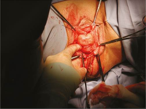
Figure 56.2 Enterocele in the posterior compartment. Reproduced with permission of Mr S Radley.
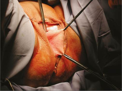
Figure 56.3 Rectocele.
Reproduced with permission of Mr S Radley.