Miscarriage
Definition
Miscarriage is defined as the spontaneous loss of pregnancy before the age of fetal viability. The World Health Organization (WHO) defines miscarriage as expulsion or extraction of a fetus or an embryo weighing 500 g or less from the mother's womb before 20 weeks of pregnancy.
The criterion for viability of pregnancy varies from 24 weeks' gestation in the United Kingdom to 28 weeks' gestation in most developing countries. Spontaneous loss of pregnancy up to 12 weeks' gestation is referred to as an early miscarriage while that between 12 to 24 weeks is termed as a late miscarriage.Epidemiology
Early miscarriage is one of the commonest complications of pregnancy affecting up to 20% of clinical pregnancies (confirmed by ultrasound scan) and early pregnancy loss is responsible for about 50,000 inpatient admissions in the United Kingdom per annum (1). The chance of a subsequent successful pregnancy following one early miscarriage is over 95% and in women with three consecutive miscarriages is over 70% (2).
Aetiology
The most common cause of miscarriage is abnormal development of the embryo or fetus (3). This may be caused by a defect in the number of chromosomes (aneuploidy) or a structural defect in one or many of the chromosomes. Such abnormalities increase with advancing maternal age. Maternal factors which increase the risk of miscarriage include antiphospholipid syndrome (APS), systemic medical disorders (such as uncontrolled diabetes, thyroid disorders, and connective tissue disease), or acute infections during pregnancy. Uterine causes of miscarriage include abnormalities such as cervical insufficiency, submucous fibroids, bicornuate uterus, septate uterus, or other Mullerian abnormalities.
The presence of parental chromosomal translocations and intrauterine adhesions due to previous uterine instrumentation or endometritis are other less common causes of miscarriage.
Pathology
Histological changes associated with miscarriage include haemorrhage into the decidua basalis and necrotic changes in the adjacent tissues. Placental villi appear thick and oedematous. Changes associated with maceration may be noted in fetal tissue.
In cases of pregnancy of unknown location (where there is no sign of an intrauterine pregnancy, ectopic pregnancy, or retained products of conception in the presence of a positive pregnancy test or serum human chorionic gonadotropin (hCG) concentration >5 IU/L), if uterine curettage is performed as part of management, it is vital to establish histological evidence of chorionic villi in the tissue obtained to exclude the possibility of an ectopic pregnancy.
The Arias-Stella reaction is a benign change in the endometrium associated with the presence of chorionic tissue and may provide the initial histological ‘clue' of an ectopic pregnancy. It is a glandular change as a physiological response to the presence of chorionic tissue. The morphological features of the Arias-Stella reaction include nuclear enlargement up to three times normal size and nuclear hyperchromasia, often accompanied by abundant vacuolated cytoplasm (4). The cells typically are stratified and the nuclei hobnail-shaped, bulging into the gland lumen. These large nuclei may contain prominent cytoplasmic invaginations. The degree and extent of the Arias-Stella reaction are highly variable in normal and abnormal intrauterine gestation, in ectopic pregnancy, and in gestational trophoblastic disease. This change occurs as early as 4 days after implantation, although it generally is seen after about 14 days (4).
Assessment of women with a possible early pregnancy loss
Women suspected to have an early pregnancy loss should be cared for in a dedicated outpatient early pregnancy assessment service. Early pregnancy assessment units (EPAUs) offer the advantages of an efficient and high-quality clinical service for women with possible early pregnancy loss, along with significant cost benefits.
A systematic approach to management of early pregnancy loss can avoid hospital admissions in 40% of cases and reduce the length of hospital stay in a further 20% (2, 5). Early pregnancy loss is associated with considerable emotional distress and appropriate support and counselling should be offered to all women (1).Diagnosis
Transvaginal pelvic ultrasound examination is the mainstay in the diagnosis of early miscarriage. Diagnosis is based on a combination of the clinical presentation correlated with ultrasound scan findings. If a transvaginal ultrasound scan is unacceptable to the woman, a transabdominal ultrasound scan should be offered and limitations of this method of scanning explained. Women should be informed that the diagnosis of miscarriage using one ultrasound scan cannot be guaranteed to be 100% accurate and there is a small chance that the diagnosis may be incorrect, particularly at very early gestational ages (1).
Threatened miscarriage
A transvaginal ultrasound scan shows a viable pregnancy in a woman presenting with cramping pelvic pain and/o r vaginal bleeding or spotting. On speculum examination, the cervix appears closed. Some of these women will progress to inevitable miscarriage regardless of the treatment offered. The best predictor of a pregnancy that will continue to viability is the presence of fetal cardiac activity (6). Intrauterine haemorrhages are commonly observed features on ultrasound examinations, especially among patients with clinically evident bleeding in early pregnancy, and the incidence has been reported to be 4-22% (7). Subchorionic haematomas usually appear as hypoechoic or anechoic crescent-shaped areas on ultrasonography (Figure 38.1). A meta-analysis suggested that the presence of subchorionic haematomas increases the risk of early or late pregnancy loss by twofold (8).
Complete miscarriage
The woman presents with vaginal bleeding and passage of tissue va- ginally (products of conception) along with abdominal pain.
The bleeding and pain appear to be settling down. On ultrasound examination, the uterine cavity is empty with a thin endometrium and a vaginal examination confirms the cervix to be closed.Incomplete miscarriage
The woman presents with active vaginal bleeding, passage of products of conception, and abdominal pain. On ultrasound examination, the uterus has evidence of retained products of conception within the uterine cavity (Figure 38.2). Ultrasound features of retained products of conception include a heterogeneous appearance and the presence of irregular tissues with or without a gestational
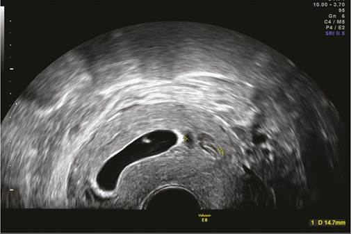
Figure 38.1 Subchorionic haematoma (marked in yellow) next to intrauterine gestational sac on transvaginal ultrasound.
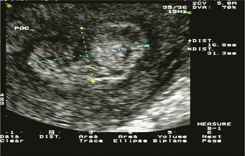
Figure 38.2 Retained products of conception (marked in yellow) within uterine cavity on transvaginal ultrasound.
sac (>15 mm diameter). The midline echo is usually disrupted or distorted.
Missed miscarriage or early pregnancy loss
This term is used when the fetus/embryo has died but is retained in the uterus for a variable period of time without symptoms of miscarriage. The woman presents with receding symptoms of pregnancy with or without vaginal bleeding or brown-coloured discharge. Transvaginal pelvic ultrasound examination confirms an intrauterine pregnancy; however, one of the following observations are made (1):
• Fetal pole with crown-rump length = 7 mm or more but no fetal heart activity (a second opinion should be sought on the viability of the pregnancy and/or a second scan performed a minimum of 7 days after the first before making a diagnosis).
• Fetal pole with crown-rump length less than 7 mm and no fetal heart activity and no change on repeat ultrasound examination performed a week later.
• Mean gestational sac diameter = 25 mm or more but no fetal pole evident (a second opinion should be sought on the viability of the pregnancy and/or a second scan performed a minimum of 7 days after the first before making a diagnosis).
• Mean gestational sac diameter less than 25 mm with no fetal pole evident and no change in scan findings after a week.
A blighted ovum is a term used for an empty gestational sac with absent embryonic pole. A possible complication of unrecognized early pregnancy loss for a prolonged time is consumptive coagulopathy leading to disseminated intravascular coagulation.
Human chorionic gonadotropin
The hCG glycoprotein is produced by the trophoblast of the implanting embryo. Circulating serum hCG concentrations are highly variable and can fluctuate widely during pregnancy. As pregnancy progresses up to 8 weeks' gestation there is a consistent doubling time over 1.94 days (2). Thereafter the doubling time lengthens and the hormone levels stabilize at about 20,000 IU/L by 10-14 weeks' gestation (9). A slower rise or declining hCG concentrations during early pregnancy may indicate an ectopic or a non-viable intrauterine pregnancy (2).
Progesterone
Serum progesterone can be a useful additional test when pregnancy of unknown location is diagnosed as its low level can predict those pregnancies that are most likely to resolve spontaneously (2).
Management
Management of possible early pregnancy loss should ideally be performed in a dedicated EPAU or acute gynaecology unit with suitably trained personnel, modern, high-quality ultrasound machines, and rapid access to serum hCG measurement. Women should be provided with appropriate psychological support and counselling. They should be provided with information about the likely impact of their treatment on future fertility and where to access support services including leaflets, web addresses, and helpline numbers for support organizations (1).
Initial management should focus on haemodynamic stabilization as well as adequate analgesia. Further management should be based on the presenting symptoms, scan findings, and serum hCG measurements. Patient choice should be encouraged at all stages and women should be helped to make informed decisions regarding their own care.
The National Institute for Health and Care Excellence guidelines in the United Kingdom recommend that expectant management for 7-14 days should be offered as the first-line management strategy for women with a confirmed diagnosis of miscarriage (1). If the resolution ofbleeding and pain indicate that the miscarriage has completed during 7-14 days of expectant management, the woman should be advised to take a urine pregnancy test after 3 weeks, and to return for individualized care if it is positive. Management options other than expectant management should be explored if (a) the woman is at increased risk of haemorrhage, (b) she has previous adverse and/or traumatic experience associated with pregnancy, (c) she is at increased risk from the effects of haemorrhage, or (d) there is evidence of infection. A repeat ultrasound scan should be offered if, after the period of expectant management, the bleeding and pain have not started (suggesting that the process of miscarriage has not begun) or are persisting and/or increasing (suggesting incomplete miscarriage).
Where clinically appropriate, women undergoing a miscarriage should be offered a choice of manual vacuum aspiration under local anaesthetic in an outpatient/clinic setting, or surgical management in a theatre under general anaesthetic.
Threatened miscarriage
This is best managed by a pelvic ultrasound examination to determine whether a fetus is present and, if so, whether cardiac activity is observed.
Over 90% of women with a live fetus will go on to deliver a baby at term and management of such women consists of reassurance and provision of emotional support (6). Hospitalization, bed rest, or administration of progestogens have not been proven to be of benefit in avoiding pregnancy loss. If the bleeding worsens or persists beyond 14 days after initial presentation, further assessment with pelvic scans should take place. If the bleeding stops, the woman should start or continue routine antenatal care.
Complete miscarriage
In cases of complete miscarriage, women should be informed of the diagnosis sensitively and provided with appropriate counselling and support. They should be offered an easy access to the EPAU in their next pregnancy.
Incomplete miscarriage
Women presenting with incomplete miscarriage should be offered the options of expectant, medical, and surgical management. In the presence of significant active bleeding, hypovolaemia should be initially corrected with crystalloids and later with compatible crossmatched blood. Prophylactic broad-spectrum antibiotics should be administered and once the patient's condition is stabilized, the remaining products of conception should be surgically evacuated from the uterus. In some patients, there may be spontaneous passage of products, thus avoiding the need for surgical evacuation. Medical and expectant management should only be offered when a woman's condition is stable and in units where access to a 24-hour telephone advice line and emergency admission facilities are available.
Expectant management
If retained products of conception measure less than 15 mm and the woman is clinically stable with minimal vaginal bleeding, she may be discharged from EPAU and offered open access to the unit if required (2). If retained products of conception measure greater than 15 mm and the patient is clinically stable with minimal vaginal bleeding she may be reassured that there is a high likelihood (7996%) that the miscarriage will complete within the next 4 weeks (2, 10- 12). The woman may be allowed home with open access to EPAU and reviewed in 2 weeks' time. If the vaginal bleeding subsides subsequently and there are no signs of infection, the woman may be discharged from care (2).
Medical management
Misoprostol is the drug of choice for medical management. The success rate of medical management of incomplete miscarriage varies between 80% and 100% (13). Medical management is more likely to induce complete miscarriage than expectant management. Risks associated with expectant or medical management include retained products of conception, infection (2%), and haemorrhage requiring blood transfusion (0.1%) (2).
Surgical management (evacuation of retained products of conception)
Surgical management has a high success rate (97- 100%) but there should be awareness of the possibility of incomplete evacuation of the uterus if symptoms do not settle postoperatively. Associated risks relate to anaesthesia, infection (2%), haemorrhage requiring blood transfusion (0.1%), cervical trauma (1%), intrauterine adhesions causing partial or complete Asherman syndrome, and uterine perforation (0.1-0.2%) (1, 2, 14). Asherman syndrome is characterized by intrauterine adhesions/fibrosis which can lead to partial or complete dysfunction of the endometrium with impairment of fertility, recurrent miscarriage (RM), and/or abnormal menstrual pattern (amenorrhoea or hypomenorrhoea). A pregnant or early pregnant uterus seems to be more susceptible to developing uterine adhesions after curettage especially if complicated by associated infection (15). Risks of incomplete evacuation or of uterine perforation may be lessened by use of transabdominal ultrasound during surgical evacuation, to guide the operator to complete the procedure safely, but this strategy is yet to be subjected to a rigorous clinical trial.
A recent systematic review assessed the effectiveness, safety, and acceptability of any medical treatment for incomplete miscarriage (before 24 weeks) (16). It included 24 randomized controlled studies (5577 women). Three trials involving 335 women compared misoprostol treatment (all vaginally administered) with expectant care. There was no difference in complete miscarriage (average risk ratio (RR) 1.23; 95% confidence interval (CI) 0.72-2.10; two studies, 150 women, random effects; very low-quality evidence), or in the need for surgical evacuation (average RR 0.62; 95% CI 0.17-2.26; 2 studies, 308 women, random effects; low-quality evidence). There were few data on ‘deaths or serious complications’. For unplanned surgical intervention, there was no difference between misoprostol and expectant care (average RR 0.62; 95% CI 0.17-2.26; two studies, 308 women, random effects; low-quality evidence). Sixteen trials involving 4044 women addressed the comparison of misoprostol (seven studies used oral administration, six studies used vaginal, two studies sublingual, one study combined vaginal plus oral) with surgical evacuation. There was a slightly lower incidence of complete miscarriage with misoprostol (average RR 0.96; 95% CI 0.94-0.98; 15 studies, 3862 women, random effects; very low-quality evidence) but with the success rate high for both methods. Overall, there were fewer surgical evacuations with misoprostol (average RR 0.05; 95% CI 0.02-0.11; 13 studies, 3070 women, random effects; very low- quality evidence) but more unplanned procedures (average RR 5.03; 95% CI 2.71-9.35; 11 studies, 2690 women, random effects; low-quality evidence). The evidence from the review evidence suggests that medical treatment, with misoprostol, and expectant care are both acceptable alternatives to routine surgical evacuation given the availability of health service resources to support all three approaches (16).
Missed miscarriage
Women with missed miscarriage should also be offered the options of expectant, medical, and surgical management.
Expectant management
The success rates of expectant management vary between 25% and 85% (17). Women should be made aware of the risk of incomplete miscarriage and the need for emergency surgical uterine evacuation in case of acute bleeding, and that there is a lower risk of infection than with surgical management. Women who chose this option should be provided with easy access to the EPAU and they should be followed up within 4 weeks.
Medical management
Medical evacuation can be achieved with the use of prostaglandin analogues (gemeprost or misoprostol) with or without antiprogesterone priming (mifepristone). Misoprostol is recommended as a single dose of 800 mcg (1). Efficacy rates vary between 13% and 96%, and are influenced by several factors including type of miscarriage, gestational sac size, total dose, duration, and route of administration of prostaglandins (2). Vaginal administration is more effective with less side effects. The side effects of the medication are nausea, vomiting, diarrhoea, and mild pyrexia. The medication should be used with caution in women with history of cerebrovascular or cardiovascular disease. The complications include infection (2%) and haemorrhage requiring blood transfusion (0.1%). Women should be advised to take a urine pregnancy test 3 weeks after medical management unless they experience worsening symptoms, in which case they should be advised to return to the healthcare professional responsible for providing their medical management to consider alternative options.
Surgical management
Women undergoing surgical evacuation for missed miscarriage should have a test for full blood count and blood group and rhesus antibody testing. They should be offered screening for infections including Chlamydia trachomatis and bacterial vaginosis. Tissue obtained at the time of evacuation of retained products of conception should be examined histologically to confirm pregnancy and to exclude gestational trophoblastic disease.
A recent systematic review was conducted to compare the safety and effectiveness of expectant management versus surgical treatment for early pregnancy loss (18). It included seven randomized controlled trials with 1521 participants. The expectant-care group was more likely to have an incomplete miscarriage by 2 weeks (RR 3.98; 95% CI 2.94-5.38) or by 6-8 weeks (RR 2.56; 95% CI 1.155.69). The need for unplanned surgical treatment was greater for the expectant-care group (RR 7.35; 95% CI 5.04-10.72). The mean percentage needing surgical management in the expectant-care group was 28%, while 4% of the surgical-treatment group needed additional surgery. The expectant-care group had more days of bleeding (mean difference 1.59; 95% CI 0.74-2.45). Further, more of the expectant-care group needed transfusion (RR 6.45; 95% CI 1.2134.42). Diagnosis of infection was similar for the two groups (RR 0.63; 95% CI 0.36-1.12), as were results for various psychological outcomes. The authors concluded that expectant management led to a higher risk of incomplete miscarriage, need for unplanned (or additional) surgical emptying of the uterus, bleeding, and need for transfusion. Risk of infection and psychological outcomes were similar for both groups. Costs were lower for expectant management. There was lack of clear superiority of either approach (18).
Sepsis associated with miscarriage
This requires urgent hospitalization and prompt institution of parenteral broad-spectrum antibiotic therapy. Once infection is controlled, careful evacuation of the uterus should be performed preferably under ultrasound guidance.
Anti- D rhesus prophylaxis should be offered at a dose of 250 IU (50 mcg) to all rhesus- negative women who have a surgical procedure to manage a miscarriage (1). Anti-D rhesus prophylaxis should not be offered to women who receive solely medical management for a miscarriage or have threatened or complete miscarriage (1).