Multifetal Pregnancy Reduction
Simi Gupta Talati and Andrei Rebarber
The Facts
The triplet and higher-order multiple birth rate was 101.6 per 100,000 births in 2017. This rate is not significantly different from 2016; however, 2016 and 2017 had the lowest triplet and higher-order multiple birth rates in more than two decades.
On the other hand, the twin birth rate was 33.3 per 1,000 births in 2017, which is only slightly less than the highest reported rate of 33.9 per 1,000 births in 2014.1Multiple gestations have been associated with a number of adverse pregnancy outcomes. Fetal mortality is increased due to a higher rate of stillbirth, and neonatal mortality is increased primarily due to a higher rate of preterm birth. Furthermore, multiple gestations have an increased risk of short-term and long-term neonatal and infant mortality switch to morbidity than singletons born at the same gestational age. Maternal morbidity is also increased with higher rates of hyperemesis gravidarum, gestational diabetes, anaemia, haemorrhage, caesarean section, post-partum depression and hypertension. Specifically, hypertension rates are 12.7% in twins and 20% in triplets as compared to 6.5% in singleton pregnancies and are associated with iatrogenic preterm delivery and abruption.2
Multifetal pregnancy reduction (MFPR) is one option to reduce the number of fetuses in a pregnancy. In this procedure, the fetus or fetuses chosen for reduction are determined based on technical considerations and the ease of accessibility for the procedure or by chorionicity in a triplet or higher-order multiple pregnancy with one or more monochor- ionic fetuses. Selective termination (ST) is the application of the same procedure as with MFPR but the fetus or fetuses chosen for reduction are based on an anomaly.
Multifetal pregnancy reduction has been shown to reduce the risk of preterm delivery and other adverse pregnancy outcomes in multiple gestations with low procedure-related loss rates.
The most recent data show that procedure-related loss rates are highest with higher starting fetal numbers (11% at five or more starting fetuses compared to 2.1% with two starting fetuses) and higher with a finishing twin pregnancy (5.3%) compared with a finishing singleton pregnancy (3.8%). Furthermore, gestational age at delivery and birthweight are lower with a finishing twin pregnancy compared with a finishing singleton pregnancy.3In pregnancies with separate chorionicities, the standard technique is to first map the location of each fetus and placenta and confirm which fetus or fetuses will be reduced (see Figure 4.1). In MFPR, the fetus located closest to the fundus or anterior abdominal wall is usually chosen for reduction due to ease of accessibility. The next step is to administer potassium chloride into the fetal heart or thorax using a spinal needle under real-time ultrasound guidance. We use 2-10 cc of 2 mEq/ml of potassium chloride with a 20-gauge
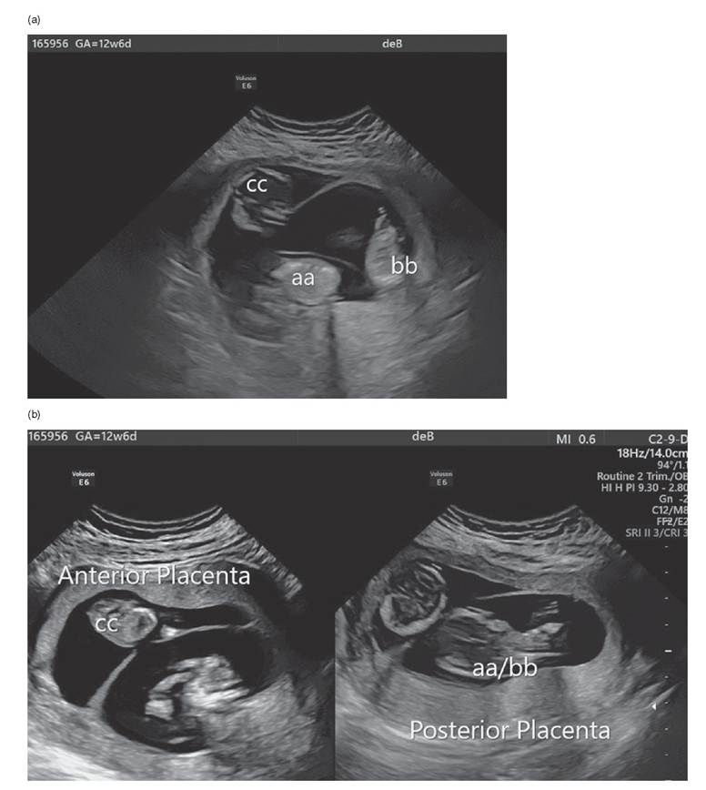
Figure 4.1 Mapping location and placentation prior to MFPR in a dichorionic-triamniotic triplet gestation
spinal needle. Asystole is usually confirmed within minutes. For additional fetuses, one may use the same needle puncture or a separate puncture depending on location.
The standard timing of the procedure is between 10 and 14 weeks gestation for MFPR and soon after diagnosis and decision-making in ST. Early embryo reduction via a transvaginal approach between 6 and 8 weeks gestation has been evaluated. However, recent data have shown a higher rate of procedure-related fetal loss (7.2% with embryo reduction versus 0.9% with fetal reduction at 11+ weeks, p = 0.039). This also translated to higher rates of overall fetal loss during the pregnancy and higher rates of miscarriage prior to 24 weeks with embryo reduction versus fetal reduction, without any decrease in the risk of preterm delivery or other adverse pregnancy outcomes.4 Therefore, fetal reduction after 10 weeks gestation is recommended for MFPR.
For selective reduction, studies have evaluated the risks and benefits of later gestational age at reduction. A large study showed a higher rate of pregnancy loss in patients undergoing ST after 18 weeks compared to less than 18 weeks (12% vs 0%, p = 0.05) and a higher rate of preterm birth with later gestational ages at ST.5 Though the dividing gestational age threshold varies between studies, most studies show lower loss rates and improved pregnancy outcomes with earlier gestational ages at ST. One study evaluated outcomes in patients undergoing ST in the second versus the third trimester and found higher rates of extreme and early preterm delivery in patients undergoing ST in the second trimester compared to the third trimester (25% vs 0%, p = 0.02). They also found no difference in pregnancy loss rates between the two groups. While this study was small, and ethical and legal considerations may prevent ST at certain later gestational ages, this study does lend some data supporting the safety of ST at later gestational ages.6Genetic screening or testing and first-trimester anatomy survey should be offered prior to MFPR in order to decrease the risk of reducing a genetic or congenital normal fetus or fetuses and continuing the pregnancy of a genetic or congenitally abnormal fetus. Chorionic villus sampling (CVS) prior to MFPR has not been shown to increase the pregnancy loss rate after MFPR and data suggest that it may actually decrease loss rates by improved selection of abnormal fetuses for reduction. One study showed a 3.1% rate of abnormal fluorescence in situ hybridisation (FISH) results in patients with a sonographically normal fetus prior to reduction.7 For patients who do not desire invasive testing, screening can be offered through ultrasound for early anatomy, nuchal translucency, nasal bone and first-trimester biochemical screening, and/or non-invasive prenatal screening. Abnormal results may then prompt patients to pursue CVS prior to MFPR or to reduce the suspected abnormal fetus.
The crosssampling rate of CVS (contamination of villi from the other fetus) reported in the literature is usually based on cases where incorrect gender determination or XX/XY mosaicism is identified. Initial sampling error rates reported as high as 4-5% have decreased to almost none.8 DNA fingerprinting can be used to determine zygosity when required in same-sex twins to determine if sampling error may have occurred. However, little published data have been described to that level of accuracy to identify the true rate of cross-sampling error in multichorionic gestations prior to fetal reduction, and this technique would not differentiate if sampling error occurred in monochorionic gestations or if monozygotic fetuses exist within multichorionic gestations.Monochorionic Multiple Gestations
In pregnancies that share a placenta (monochorionic), potassium chloride injections into the fetal heart or thorax cannot be performed because of concern about transplacental passage of the potassium chloride to the other fetus(es)and because hemodynamic changes in the reduced fetus may cause neurological morbidity or mortality to the other fetus. Therefore, an alternative technique, primarily some form of umbilical cord occlusion, is utilised.
There are four main techniques for umbilical cord occlusion: suture ligation, laser coagulation, bipolar coagulation and radiofrequency ablation. In the original suture ligation procedure, a port is introduced into the uterine cavity and a vicryl suture is placed into the cavity through the port. The suture is then looped around the cord by ultrasound guidance and tied in a knot extra-corporeally, and a knot pusher is used to secure the knot. This is repeated in a second position along the cord to ensure complete cord occlusion.9 In the technique of laser coagulation, again a small port is used for uterine entry, and an operative endoscope is used to visualise the appropriate cord. The umbilical vessels are then photocoagulated using a 600 microm quart Nd-YAG noncontact laser fibre (with a power of 20-40 W) through the operating channel of the endoscopy.
Each pulse lasts 2-3 seconds and is repeated until coagulation is completed as determined by lack of blood flow using colour and pulsed Doppler ultrasound. If bipolar coagulation is being used, the umbilical cord is grasped with a 3 mm bipolar forceps introduced through a uterine port and coagulation is performed at a power setting of 30-50 watts for 30 seconds. Additional cord segments can be coagulated as a precaution and cessation of flow in the umbilical vessels is confirmed by colour Doppler. In the technique of radiofrequency ablation, a 17-gauge Starburst radiofrequency needle is inserted into the fetal abdomen adjacent to the area of the umbilical cord insertion and the prongs of the device are partially deployed. Radiofrequency energy is applied for 4 minutes at 95 watts to generate a target temperature of 100 degrees Celsius. The procedure is repeated for an additional one or two cycles until cessation of cord blood flow is confirmed using colour and power Doppler velocimetry.There are pros and cons to the different procedures which may guide choice of procedure. Bipolar coagulation requires a larger-calibre operative sleeve and therefore may be more useful in mid second-trimester cases where there is enough amniotic fluid to accommodate the larger operative sleeve or where additional procedures may be required through the port. The benefit of bipolar coagulation is complete occlusion with first application. Radiofrequency or laser ablation has the benefit of a smaller-diameter device and therefore may be preferred in cases at earlier gestation, with low amniotic fluid or with a shorter umbilical cord. However, the occlusion is slower, which may alter flow patterns between the fetuses and increase the risk of fetal demise. There is also a concern about an increased risk of rupture of membranes from thermal damage from the device. Finally, suture ligation may be the best option at late gestational ages when the bipolar forceps may not fit around the umbilical cord.
In recent years, only bipolar coagulation and radiofrequency ablation have been compared in observational trials and a large systematic review and meta-analysis showed no difference in co-twin death rates, live birth rates, neonatal death rates or overall survival. There was a slight increase in preterm premature rupture of membranes with bipolar coagulation versus radiofrequency ablation (28.2% vs 17.7%, p = 0.01), but no difference in preterm delivery rates before 28 or 32 weeks of gestation. This study also showed that the outcome data with selective reduction in monochorionic twin pregnancies are worse than with dichorionic twin pregnancies with an overall survival rate - live birth rate minus neonatal death rate - of only 76.8% with radiofrequency ablation and 79.1% with bipolar coagulation. The risk of preterm delivery was also high with a mean gestational age at delivery of 34.7 weeks with radiofrequency ablation and 35.1 weeks with bipolar coagulation.10 The second, older systematic review also showed lower survival rates in selective reduction in monochorionic pregnancies of 86% with radiofrequency ablation, 72% with bipolar coagulation, 72% with laser coagulation and 70% with cord ligation.11 The third study looked at long-term neurodevelopmental impairment in children at least two years of age after selective reduction in monochorionic pregnancies and found a 6.8% risk of neurodevelopmental impairment.12
Most selective reduction procedures in monochorionic pregnancies occur in the second trimester soon after the diagnosis for need is made. One systematic review showed improved survival rates when the procedure was performed after 18 weeks (89% vs prior to 18 weeks 69%, p = 0.02). However, the second study looking at timing of intervention of twin-reversed arterial perfusion (TRAP) cases showed worse outcomes when cases were managed conservatively with serial ultrasound, and therefore some advocate for early intervention in the second trimester as soon as the diagnosis is made.13
In addition to the different techniques for cord occlusion, some authors recommend antibiotics prior to, during and post procedure. Some also use tocolytics such as nifedipine and/or indomethacin around the time of the procedure. Depending on the author and procedure, patients are either discharged home the same day or monitored in the hospital for 24-48 hours prior to discharge. All patients are followed up at regular at 1-2-week intervals and most receive either a detailed fetal neurosonogram or fetal MRI to evaluate for cerebral injury post procedure.
Multifetal pregnancy reduction is usually not recommended in monochorionic- monoamniotic twin pregnancies due to the residual risk of cord entanglement even with cord occlusion. However, there have been reports in cases of fetal anomalies and the most recent case report discussed a technique of bipolar cord occlusion followed by laser ablation to transect the cord in two cases with selective intrauterine fetal demise of one twin. Both cases were reported to have good outcomes.14
The Issues
Multifetal Pregnancy Reduction in Triplet and Twin Pregnancies
While pregnancy loss rates continue to be low after reduction of trichorionic-triamniotic (TT) and dichorionic-diamniotic (DD) pregnancies, the reduction in adverse pregnancy outcomes appears to be more modest. For example, a recent study comparing continued TT pregnancies to TT pregnancies reduced to DD pregnancies showed no difference in preterm delivery rates prior to 34 weeks, but did show an increase in birthweight and a decrease in caesarean sections with MFPR. This study also showed no difference in pregnancy loss rate prior to 24 weeks between ongoing TT pregnancies and MFPR to DD pregnancies.15
Another study comparing continued DD pregnancies with MFPR to a singleton pregnancy showed reduction in preterm delivery rates prior to 37 weeks and 34 weeks with MFPR, but no reduction in preterm delivery rates prior to 32 weeks. It also showed a decrease in birthweight less than 10% and 5%, pre-eclampsia and caesarean section with MFPR. This study showed no difference in unintended pregnancy loss rates between DD and MFPR to singleton pregnancies.16 While the most dramatic survival benefits are seen with reductions from higher initial starting numbers of fetuses in higher-order multifetal pregnancies, limited data have been published to identify whether there is improved longterm outcomes in twins reduced to singletons given the advances in neonatal care of late preterm births.
In dichorionic-triamniotic (DT) triplet pregnancies, there are three options for a triplet pregnancy of mixed chorionicity: reduction to two fetuses by reducing one of the mono- chorionic pair, reduction to two fetuses by preserving the monochorionic pair, and reduction to one fetus by reducing both fetuses from the monochorionic pair. The reduction of the monochorionic-diamniotic (MD) pair or the fetus with a separate placenta increases the gestational age at delivery compared to expectant management of DT pregnancies. However, the pregnancy loss rate appears to be higher with reduction of the fetus with a separate placenta, but similar between reduction of the MD pair and ongoing DT pregnancies. Overall reduction of one or more fetuses significantly reduces the risk of severe prematurity with a marginal impact on the miscarriage rate.
Management of Pregnancies after Multifetal Pregnancy Reduction or Selection Termination
While more recent studies have focused on comparing pregnancy outcomes between ongoing multiple gestation pregnancies and pregnancies after reduction, older studies compared pregnancy outcomes in pregnancies after reduction and pregnancies that originated as a singleton pregnancy. In these older studies, adverse pregnancy outcomes after reduction were higher than in pregnancies that originated as singleton pregnancies. Specifically, these older studies showed high rates of preterm delivery and intrauterine growth restriction in pregnancies after reduction than in pregnancies that originated as a singleton pregnancy. Therefore, one would assume that pregnancies after reduction would require a higher level of surveillance than routine singleton pregnancies.
Few studies have reviewed surveillance strategies after fetal reduction. Our group assessed the validity of vaginal fetal fibronectin (fFN) as a screening test for spontaneous preterm delivery in asymptomatic patients who had undergone MFPR. In this cohort of 63 patients, 13 singleton gestations and 50 twin gestations, a median of 4 fFN assays were performed per patient. A total of 234 fFN tests were performed with 222 negative results and 12 positive results. The fFN test had similar validity to predict spontaneous preterm birth in these at-risk pregnancies as previously published cohorts.17 Additionally, our group also evaluated the utility of serial cervical-length evaluation in asymptomatic twins with and without fetal reduction procedures to address the potential effect from the procedure on the measurements. The study group of 35 twins after MFPR was compared to 83 twin gestations who had not had the procedure, and the results noted that cervical length across gestation was not affected by the MFPR procedure.18 However, there are no studies evaluating prospectively different management protocols after reduction. Our management suggestions are listed in what follows.
Management Options
Multifetal Pregnancy Reduction or Selective Termination in Pregnancies with Separate Chorionicities
Pre-pregnancy Counselling
• Patients are counselled about the risks and benefits of MFPR or ST based on starting number, ending number and gestational age at decision-making.
• Patients who are planning on undergoing MFPR are counselled about the options of genetic screening/testing and are offered non-invasive prenatal screening, nuchal translucency/nasal bone screening and/or CVS with FISH, karyotype and/or microarray. They are also offered first-trimester anatomy screening to assess for congenital anomalies.
• Patients are scheduled for MFPR as soon after genetic screening/testing results are returned or for ST as soon as the diagnosis of a fetal anomaly is made, but not before 10 weeks of gestation.
Multifetal Pregnancy Reduction or Selective Termination Protocol
• Patients undergo ultrasound mapping of each fetus and placenta. For patients undergoing genetic screening/testing, all attempts are made to have the same physician and sonographer who perform the mapping for genetic screening/testing also perform the reduction procedure in order to minimise the risk of error.
• Two to 10 cc of 2 mEq/ml of potassium chloride are transabdominally administered into the fetal heart or thorax with a 20-gauge spinal needle under ultrasound guidance. Asystole is confirmed. For additional fetuses, one may use the same needle puncture or a separate puncture depending on location of the other fetus(es).
• Rhogam is administered as needed for Rh-negative patients.
Post-reduction Pregnancy Management
• Maternal serum alpha feto-protein levels are not drawn for patients who have undergone reduction procedures as they may be falsely elevated.
• Patient instead undergoes an initial anatomy scan at 16-18 weeks to evaluate for congenital anomalies. They also undergo the routine anatomy scan at 20-22 weeks.
• Patients are followed with serial cervical length measurements to assess for risk of preterm delivery. Low-risk treatment options for short cervix such as vaginal progesterone for patients with a cervical length < 2.5 cm are recommended in appropriate patients, while treatment options such as cervical cerclage are offered on an as-needed, case-by-case basis, particularly in the setting of an extremely shortened or dilated cervix.
• Patients are followed with serial ultrasounds for fetal growth every 4 weeks after 20 weeks to detect intrauterine growth restriction. Additional monitoring for pregnancies diagnosed with intrauterine growth restriction such as antenatal testing with biophysical profiles or fetal Doppler evaluation is performed as needed.
• Routine antenatal testing with biophysical profiles is started weekly at 32-34 weeks of gestation or earlier if clinically indicated.
• Timing and mode of delivery are per usual obstetric indications.
Selective Termination in Monochorionic Pregnancies
• Patients undergo an initial anatomy scan at 16-18 weeks to evaluate for congenital anomalies. They also undergo a routine anatomy scan at 20-22 weeks.
• Patients are followed with serial cervical length measurements to assess the risk of preterm delivery. Low-risk treatment options for short cervix such as vaginal progesterone for patients with a cervical length < 2.5 cm are recommended in appropriate patients, while treatment options with higher risks such as cervical cerclage are offered as needed on a case-by-case basis, particularly in the setting of an extremely shortened cervix or a dilated external observational study prior to 24 weeks.
• Patients are followed with serial ultrasounds for fetal growth every 4 weeks after 20 weeks to detect intrauterine growth restriction. Additional monitoring for pregnancies diagnosed with intrauterine growth restriction such as antenatal testing with biophysical profiles or fetal Doppler evaluation are performed as needed.
• Timing and mode of delivery are per usual obstetric indications.
Key Points
• Obstetric caregivers should be aware that multifetal pregnancies increase maternal and perinatal morbidity and mortality. The greater the number of fetus(es) the higher the risks to both mothers and infants.
• Non-directive patient counselling should be offered to all women with multiple gestations regarding the potential medical benefits of fetal reduction.
• Prior to fetal reductions, attempts should be made with either non-invasive or invasive testing to determine the genetic/structural normality of the fetus that will remain.
• Multifetal pregnancy reduction and selective termination are generally performed between 11 and 14 weeks or as soon as possible after diagnosis of an anomaly, and the loss rate of the pregnancy is based on the starting number as well as the number remaining.
• Biochemical screening for aneuploidy and/or spina bifida should not be performed after the procedure.
• Serial assessment of fetal growth should be considered in these pregnancies, particularly in remaining twin gestations.
• Consider second-trimester cervical length screening +/- fFN screening in both singleton and twin gestations.
References
1. Martin JA, Hamilton BE, Osterman MJK, Driscoll AK, Drake P. Births: final data for 2017. National Vital Statistics Reports 2018;67(8):1-50.
2. American College of Obstetricians
and Gynecologists. Multifetal gestations: twin, triplet, and higher-order multifetal pregnancies. Practice Bulletin No. 169. Obstet Gynecol 2018;128: e131-e146.
3. Stone J, Ferrara L, Kamrath J, Getrajdam J, Berkowitz R, Moshier E et al. Contemporary outcomes with the latest 1000 cases of multifetal pregnancy reduction (MFPR). Am J Obstet Gynecol 2008;199:406.e1- 406.e4.
4. Kim MS, Choi DH, Kwon H, Ahn E, Cho HY, Baek MJ et al. Procedural and obstetrical outcomes after embryo reduction vs fetal reduction in multifetal pregnancy. Ultrasound Obstet Gynecol 2019;53:214-18.
5. Bigelow C, Factor S, Moshier E, Bianco A, Eddleman K, Stone J. Timing and outcomes after selective termination of anomalous fetuses in dichorionic twin pregnancies. Prenat Diagn 2014;34:1320-5.
6. Dural O, Yasa C, Kalelioglu I, Can S, Yilmaz G, Esmer AC et al. Comparison of perinatal outcomes of selective termination in dichorionic twin pregnancies performed at different gestational ages. J Matern Fetal Neonatal Med 2017;30(12):1388-92.
7. Rosner M, Pergament E, Andriole S, Gebb J, Dar P, Evans M. Detection of genetic abnormalities by using CVS and FISH prior to fetal reduction in sonographically normal appearing fetuses. Prenat Diagn 2013;33:940-4.
8. Evans MI, Andriole S, Evans SM. Screening and testing in multiples. Clin Lab Med 2016;36(2):289-303.
9. Wilmasundra RC. Selective reduction and termination of multiple pregnancies. Semin Fetal Neonat 2010;15:327-35.
10. Gaerty K, Greer R, Kumar S. Systematic review and metaanalysis of perinatal outcomes after radiofrequency ablation and bipolar cord occlusion in monochorionic pregnancies. Am J Obstet Gynecol 2017:637-43.
11. Rossi AC, D'Addario V. Umbilical cord occlusion for selective feticide in complicated monochorionic twins:
a systematic review of literature. Am J Obstet Gynecol 2009:123-9.
12. Van Klink JMM, Koopman HM, Middeldorp JM, Klumper FJ, Rijken M, Oepkes D et al. Long-term neurodevelopmental outcomes after selective feticide in monochorionic pregnancies. BJOG 2015;122:1517-24.
13. Bebbington M. Selective reduction in complex monochorionic gestations. Am J Perinatol 2014;21:S51-S58.
14. Greimel P, Csapo B, Haeusler M, Lang U, Klaritsch P. A modified technique for cord transection in monochorionic monoamniotic twin pregnancies. Fetal Diagn Ther 2018;44:236-40.
15. Herlihy N, Naqvi M, Romero J, Gupta S, Monteagudo A, Rebarber A et al. Multifetal pregnancy reduction of trichorionic triplet gestations: what is the benefit? Am
J Perinatol 2017;34:1417-23.
16. Viera L, Warren L, Pan S, Ferrar L, Stone J. Comparing pregnancy outcomes and loss rates in elective twin pregnancy reduction with ongoing twin gestations in a large contemporary cohort. Am J Obstet Gynecol 2019;221:253.e1-8.
17. Roman AS, Rebarber A, Lipkind H, Mulholland J, Minior V, Roshan D. Vaginal fetal fibronectin as a predictor of spontaneous preterm delivery after multifetal pregnancy reduction. Am
J Obstet Gynecol 2004;190(1):142-6.
18. Rebarber A, Carreno CA, Lipkind H et al. Cervical length after multifetal pregnancy reduction in remaining twin gestations.Am J Obstet Gynecol 2001; 185(5):1113-17.