Placenta accreta (morbidly adherent placenta)
Definition
Placenta accreta broadly refers to abnormal placentation of varying degrees of morbid adherence placenta, subclassified into accreta, increta, and percreta based on the degree of penetration of chorionic villi into the uterine wall.
It probably results from defective decidualization of the implantation site or implantation in a defective uterine scar (31, 42). In placenta accreta, the chorionic villi are attached to the myometrium instead of the decidua, while in percreta it penetrates into the myometrium and in percreta, the placenta penetrates through the myometrium into the serosal layer and surrounding viscera, commonly the bladder. Accreta is used as an umbrella term for the spectrum of accreta-increta-percreta in this chapter. Increasingly, the term ‘morbidly adherent placenta' is being used to encapsulate the various forms of this type of placenta.Incidence
The incidence of placenta accreta has increased progressively over the past decade in many regions of the world, an increase largely attributable to the increasing caesarean section rates across the world. Rates of up to 1 in 700 deliveries have been reported (43). In a series of histologically confirmed morbidly adherent placenta, placenta accreta accounted for 79% while placenta increta and percreta made up 14% and 7% respectively (44).
Risk factors
The most important risk factor for abnormally adherent placenta praevia is previous caesarean delivery; with risks increasing with the number of prior caesarean sections (Box 22.5). Up to 11% of placenta praevia in a previous caesarean section will be associated with placenta accreta rising to 40% in patients with three previous caesarean sections and placenta praevia (45). A history of prior caesarean section in the absence of placenta praevia is also associated with an increased risk of placenta accreta (46).
Box 22.5 summarizes the risk factors for placenta accreta.Diagnosis
Most cases of placenta accreta are suspected after the routine midtrimester ultrasound scan or following episodes of bleeding or persistent haematuria in cases of placental extension into the bladder. Transabdominal (Figure 22.3) and transvaginal ultrasound assessment of the placenta in combination with colour Doppler remains the most reliable tool for the diagnosis of abnormally invasive placenta (47-48). Sonographic features of abnormal placenta invasion include the presence of lacunae or venous lakes, loss or disruption of the retroplacental hypoechoic (sonolucent) zone, exophytic mass extending through the serosa, and bulging of the placenta into the posterior wall of the bladder (49). In addition, colour Doppler is highly suggestive of placenta accreta when there is abnormal lacunar flow with prominent vessels over the peripheral subplacental area and hypervascularity of the serosa-bladder interface. The role of magnetic resonance imaging (MRI) in the diagnosis of abnormal placental invasion has been extensively reviewed and found to be comparable to ultrasonography and colour Doppler though with a slightly lower accuracy before 24 weeks' gestation (50-52). MRI is not recommended as the primary diagnostic modality but it has a complementary role where ultrasound findings are inconclusive, in posterior placentas, and to assess the depth or myometrial invasion (53). The MRI features suggestive of placenta accreta include:
• abnormal placenta vascularity
• uterine bulging
• heterogeneous signal intensity on T2-weighted imaging
• heterogeneous signal intensity within the placenta mass
• interruption of the myometrium.
Though not frequently used, three-dimensional ultrasonography has been used in the diagnosis of placenta accreta using
Box 22.5 Risk factors for placenta accreta
• Previous caesarean section with placenta praevia
• Previous uterine scarring or surgery:
— Myomectomy
— Hysteroscopic resection
— Adhesiolysis
— Endometritis
— Uterine curettage
— Endometrial ablation
• In vitro fertilization
• Caesarean section scar pregnancy
• Maternal age over 35 years
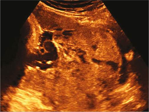
Figure 22.3 Ultrasound diagnosis of placenta accreta—invasive placenta with irregular lacunae.
key diagnostic features such as increased vascularity of the serosa-bladder interphase and irregular tortuous intraplacental vascularization (54).
Timely confirmation of the diagnosis of placenta accreta is crucial to allow sufficient time for advance planning of delivery (Box 22.6). Following the initial ultrasound diagnosis, a further follow-up scan should be performed in the third trimester around 32 weeks in stable patient. An MRI is typically performed at this gestation as well and any additional information gained could be useful in planning the delivery.
Management
The antenatal management of patients with placenta accreta follows the same principles as described for placenta praevia. The distinctive difference, however, is the need for detailed and multidisciplinary advanced planning based on the anticipated complexity of the case. Preoperative planning using the care bundle itemized in Box 22.4 is recommended in all cases of placenta accreta.
Preterm delivery should be anticipated in patients with placenta accreta and a low threshold for administering antenatal corticosteroids to enhance fetal lung maturity should be adopted in symptomatic patients after 24 weeks of gestation.
Box 22.6 Complications of placenta accreta
• Massive haemorrhage
• Unscheduled hysterectomy
• Preterm delivery (spontaneous and iatrogenic)
• Massive transfusion
• Disseminated intravascular coagulopathy and adult respiratory distress syndrome related to massive transfusion
• Sepsis
• Spontaneous uterine rupture
• Intensive care unit admission
• Maternal mortality
Delivery
The main underlying principle with respect to the timing of delivery is the avoidance of emergency delivery; therefore, elective delivery by caesarean section is recommended at 35-36 weeks of gestation. Earlier delivery at 34 weeks should be considered in patients with recurrent symptoms especially those with placenta percreta that involves adjacent viscera.
The plan for delivery should involve senior clinicians from the relevant specialties including obstetrics, anaesthesia, radiology, and urology and a gynaecological oncologist surgeon and neonatolo- gists. The components of the care bundle could be adapted to the setting based on the skill set and resources available. There should be a comprehensive discussion regarding the intended surgical approach and this should be explained carefully to the patient and documented in her records.
Surgical management
The surgical options for placenta accreta include the following:
• Delivering the baby through an incision away from the site of the placenta, leaving the placenta undisturbed, closing the uterus, and proceeding to a caesarean hysterectomy. This option has been associated with reduced morbidity and blood loss and is ideally recommended in those patients in whom future fertility is not a priority (31).
• Delivering the baby through an incision away from the site of the placenta, leaving the placenta undisturbed, then trimming the umbilical cord and closing the uterus with the placenta in situ. The patient is kept for an initial period of monitoring with antibiotics cover and assessment of bleeding. This is followed up by a prolonged period of follow-up with beta-human chorionic gonadotropin and serial ultrasound scans until the placenta mass completely resolves. This is an option in those patients who wish to preserve their uterus. During the follow-up period, this conservative approach is associated with a significant risk of infection and sudden heavy bleeding that would necessitate an emergency caesarean hysterectomy. The role of methotrexate in enhancing the breakdown of placenta trophoblastic tissue is debatable with no clear evidence of benefit and significant drug related morbidity, hence routine use is not advised (55-56).
• Delivering the baby followed by excision of the placenta bed with or without separating the placenta, followed by uterine reconstruction.
This option can be associated with significant haemorrhage and adequate blood and blood products must be available. The excisional approach is usually appropriate when there is a clear margin of normal myometrium below the lower edge of the placenta, typically in focal accreta (55). Recently a triple P procedure has been described which is along the same principles as this approach but can retain some chorionic tissue (57)• In some cases, especially those with bladder involvement, a joint procedure with urologists is advised and the placement of ureteral stents could be of benefit in selected cases to assess the integrity of the urinary tract, particularly during caesarean hysterectomy.
There is a significant risk of haemorrhage when the placenta partially separates and speedily adjunct surgical measures have to be taken to stop bleeding. These measures include internal iliac artery ligation, balloon tamponade, compression sutures (e.g. B-Lynch sutures), and a timely resort to hysterectomy if bleeding persists. The use of a prophylactic catheter placement for balloon occlusion or embolization has become part of the preoperative plan in many centres, though with variable evidence of clear benefit (58). The catheter, if left in situ, can be useful in the immediate postpartum period to manage postpartum haemorrhage.
The use of cell saver technology is encouraged where the expertise and facilities are available, especially in those patients who decline donor blood transfusion.
Postoperative care
The management of patients with placenta accreta follows similar principles as previously outlined for major placenta praevia. Access to a critical care bed should be part of the preoperative workup as most patients with prolonged surgery and massive blood transfusion will require postoperative monitoring in the critical care setting in the immediate postoperative period. Careful attention should be paid to assessment of blood loss from pelvic drains, fluid balance, thromboprophylaxis, as well as antibiotic prophylaxis in those patients with placenta tissue in utero.