Screening for Fetal Abnormality in Multiple Pregnancy
Carolina Bibbo, Julian N. Robinson and Beryl Benacerraf
The Facts
Chorionicity and the Risk of Fetal Abnormality
Determination of chorio-amnionicity in the early first trimester is of great importance to establish the amount of risk that can be associated with the pregnancy.
Monochorionic twin pregnancies have an increased incidence of perinatal morbidity and mortality given the shared placental-fetal circulation. Such complications can be due to the occurrence of twintwin transfusion syndrome (TTTS) and selective intrauterine growth restriction (sIUGR) or a mixture of the two (both of these conditions are dealt with in detail in Chapter 13).1 Chorionicity and amnionicity are most accurately determined by ultrasound in the first trimester after 7 weeks with a sensitivity of more than 98%.1 The typical findings on ultrasound are the ‘lambda sign' for dichorionic-diamniotic twins and the ‘T-sign’ for monochorionic-diamniotic twins (Figures 7.1, 7.2). Monochorionic-monoamniotic twins are characterised by the absence of the inter-twin membrane (Figure 7.3).Although chorionicity can be easily determined by ultrasound, chorionicity does not accurately determine zygosity (dichorionic twins can be monozygous). Knowing the zygosity is important to determine the risk of aneuploidy for the pregnancy. Zygosity needs to be included in genetic counselling, the management of genetic screening and diagnostic testing options. Knowing the chorionicity is important for the management of these pregnancies that are at risk for TTTS and sIUGR. Chorionicity determines the protocol for routine antenatal care and ultrasound surveillance that will be followed throughout the pregnancy (ultrasound surveillance every 2-3 weeks for monochorionic twins and monthly
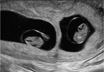
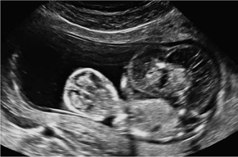
Figure 7.1 This image represents a dichorionic- diamniotic twin pregnancy at 8 weeks.
The inter-twin membrane with the 'twin peak' or 'lambda sign' is characteristic of the triangular projection of tissue from a fused dichorionic placenta.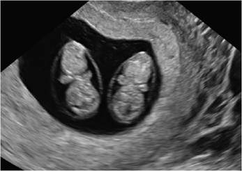
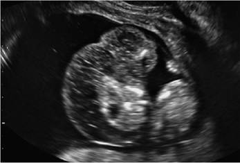
Figure 7.2 This image represents monochorionic- diamniotic twins at 9 weeks.
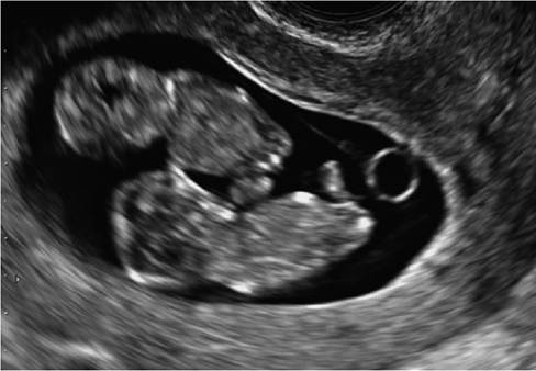
Figure 7.3 Thisimage represents monochorionic- monoamniotic twins at 11 weeks.
The inter-twin membrane is absent.
for dichorionic twins). Chorionicity, amnionicity and zygosity are dealt with in detail in Chapter 3.
Dizygotic twin pregnancies carry double the risk of aneuploidy as a maternal age- matched singleton pregnancy (with the pregnancy likely being discordant for aneuploidy). Twin conceptions can also result in a single, normal-appearing fetus adjacent to a partial or complete molar twin (Figure 7.4).
Structural anomalies are more common in monozygotic twins, at least double that of singletons. The chance of a malformation in at least one fetus is four times as high as in a singleton pregnancy (8%). This risk is highest in monoamniotic twins (17%).1 There is conjecture that the mechanical process of the embryo splitting may precipitate structural abnormalities. Dizygotic twins have the same incidence of malformations as singletons, but the overall risk of a malformation in at least one fetus is twice as high as in a singleton pregnancy (4%).2
Despite being genetically identical, monozygotic twins can be discordant for structural abnormality. Only 20% of malformations are concordant in monozygotic twins.2 Congenital heart disease is more prevalent in monochorionic twins, a proportion of which is caused in response to the abnormal physiology of TTTS.3 Among the most
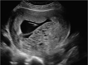
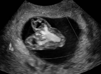
Figure 7.4 This image represents a twin conception with a normal fetus and a coexistent molar pregnancy at 9 weeks.
common structural malformations in twin pregnancies are cardiac anomalies, neural tube and brain malformations and gastrointestinal and abdominal wall defects.
A common error in clinical practice is to miss multiple structural abnormalities once one abnormality has been diagnosed. The distraction of finding an abnormality can affect the clinical performance of the rest of the study. However, detecting one abnormality should trigger increased suspicion for another anatomic defect as an underlying genetic syndrome may be present.
Some of the malformations seen in monozygotic twins are inherent to the twining process itself, such as acardiac twinning and conjoined twins. These malformations can be easily diagnosed in the first trimester. It has been our experience that the twin-reversed arterial perfusion (TRAP) sequence, also known as an acardiac twin, can be misdiagnosed as a structural abnormality or as a co-twin fetal demise in the first-trimester scan (Figures 7.5, 7.6). In these situations, colour Doppler showing reversed perfusion within the acardiac twin or the use of transvaginal ultrasound to enhance anatomic detail can aid in the diagnosis. Conjoined twins can also be diagnosed in the first-trimester scan. The typical findings are proximity of fetal poles and lack of independent movement of body/limbs from each twin. Sometimes the fusion of the organs can be apparent even in the first trimester (Figures 7.7, 7.8).
For all first-trimester ultrasound performed in this setting, the operator should have a low threshold for utilising both transabdominal and transvaginal ultrasound. In modern practice, the use of transvaginal ultrasound is routine due to higher-frequency transducers and closer proximity to the fetal parts. Doppler can be useful but should be used with caution and for specific indications due to the higher power exposure of this modality.
Discrepancy in Inter-twin Crump-Rump Length Measurement
The observation of size discordance in the first trimester is important.
When there is discordance in the crump-rump length (CRL) measurement in twins, the dating should be established by the larger. The discordance in CRL is calculated as the difference between the larger CRL and the smaller CRL, divided by the larger CRL and multiplied by 100.The prevalence of discordance in inter-twin CRL is not accurately known. The body of literature pertaining to this subject is heterogeneous not only in the definition of the discordance thresholds, but also in the inclusion criteria of the study populations (aneu- ploid fetuses, anomalous fetuses, monochorionic twins). Most published reports define inter-twin CRL discordance when the discordance in CRL is ≥10%.4 This 10% discordance
Figures 7.5 and 7.6 These images represent a monochorionic-diamniotic twin pregnancy at 12 weeks affected by twin-reversed arterialperfusion (TRAP sequence), also known as acardiac twin.
represents the 90th or 95th percentile distribution in most studies. The degree of CRL discrepancy is not related to chorionicity or the mode of conception.5 Monochorionic twins are at a much higher risk of fetal size discrepancy, loss and other complications.6
The studies that have focused on the outcomes of twins with inter-twin CRL discordance in a dichorionic twin population (excluding monochorionic twins) have conflicting findings. Kalish et al. and Salomon et al. in the early 2000s showed that dichorionic twins with inter-twin CRL discordance > 90th percentile (>10%) were more likely to have a twin with either a chromosomal or anatomic anomaly.5,7 Kalish et al.'s work emphasised the poor predictive value of this finding showing that when the inter-twin CRL discordance was >10%, the sensitivity to predict birth discordance (>20%) was 18.8% with a specificity of 92.1%.7 Fareeduddin et al.
have published that in euploid dichorionic twins that are structurally normal, first-trimester inter-twin CRL discordance is associated with adverse pregnancy outcomes, including preterm birth, preterm premature rupture of the membranes (PPROM) and lower birthweights.6 Harper et al. have reported that dichorionic twin pregnancies affected by CRL discordance are more likely to result in the loss of one of the twins prior to 20 weeks (10.5 vs 1.3%; RR 7.8 (95% CI 3-20.5)). Furthermore, their study showed that the risk of structural abnormality was 60% higher than in twins with concordant CRL. However, if the pregnancy did not result in a loss of one twin or one of the twins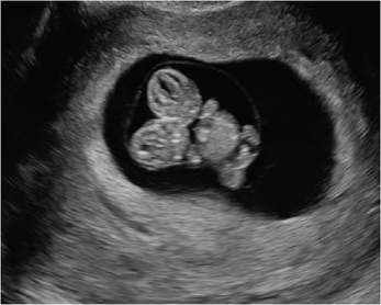
Figures 7.7 and 7.8 These images represent conjoined monochorionic-monoamniotic at 9 weeks. Colour Doppler is used to clarify the anatomy and identification of the conjoined twins which are thoracopagus (fusion from thorax to umbilicus). (A black and white version of this figure willappear in some formats. For the colour version, please refer to the plate section.)
having an anomaly, the pregnancy was not associated with a higher risk of stillbirth, intrauterine growth restriction or preterm delivery prior to 34 weeks.8
D’Antonio and his collaborators demonstrated that once structural malformations and aneuploidy have been excluded, CRL discordance is weakly associated with poor adverse perinatal outcome, and as such, it is a poor predictor of these events in both dichorionic and monochorionic twins.9 A recently published systematic review of 17 studies confirmed this finding. This review showed that inter-twin CRL discordance has been associated with a higher risk of adverse perinatal outcomes including fetal loss, weight discordance, fetal anomalies and preterm delivery.
However, despite this association, such discordance had a low positive predictive value and its use in clinical practice is limited.4 These authors suggest that such a finding should not change clinical management.In conclusion, discordance in inter-twin CRL measurement raises concern to the obstetrician. Studies including only dichorionic twins or both dichorionic and monochor- ionic have found an association with fetal loss and fetal anomaly. As fetuses with chromosomal abnormalities have been found to have a smaller CRL than expected, CRL discordance might prompt investigation for aneuploidy. Despite the potential association with poor pregnancy outcome, the CRL discordance has a poor predictive value of such events. However, if there is a large discrepancy in the size of the gestational sacs that accompanies the CRL discrepancy, the prognosis is guarded (Figure 7.9). In our opinion, first-trimester CRL discordance warrants concern for aneuploidy and structural abnormality and should trigger genetic counselling and further ultrasound surveillance. No
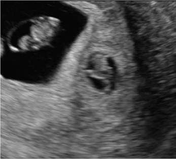
Figure 7.9 This image represents dichorionic- diamniotic twins at 7 weeks with a large discrepancy in the size of the gestationalsacs (smallgestationalsac size of the smaller twin) that accompanies the CRL discrepancy.
long-term predictions regarding pregnancy outcome should be made due to such findings without further pertinent information.
Nuchal Translucency Measurement Discordance
The nuchal translucency (NT) thickness can be measured between 10 and 13 weeks to screen for aneuploidy and fetal structural abnormality. As in singleton pregnancies, a thickened nuchal translucency or cystic hygroma is equally concerning for fetal aneuploidy and fetal abnormality in twin pregnancies. In twin pregnancies, sonographic genetic screening with nuchal translucency has an increased importance since serum screening is less accurate.
The interpretation of maternal serum genetic screening is challenging in dichorionic twin pregnancies (the majority dizygotic) as both twins contribute to the concentration of these markers and the overall results do not specify which twin is affected. The measurement of the nuchal translucency thickness can certainly improve the aneuploidy detection rate and better identify the affected twin in these pregnancies. The test sensitivity in dichorionic twins of first-trimester maternal serum markers and nuchal translucency measurement is 86% (95% CI 73-94) and the test sensitivity in monochorionic twins is 87% (95% CI 5398).10
It is important to mention that early in the first trimester, underlying hemodynamic changes in TTTS can result in an increased NT thickness in the recipient twin.11 Therefore the false positive rate of NT screening is higher in monochorionic twins than in dichorionic twins, as the pathogenesis can reflect early severe TTTS rather than aneuploidy.
Kagan et al. studied inter-twin discordance in NT and its predictive value in severe TTTS. Their study excluded pregnancies diagnosed with chromosomal or major structural abnormalities. They found that 25% of monochorionic twins had an inter-twin discordance in NT of ≥20%. When there was such a finding, the risk of complications (death of one or both twins < 18 weeks or severe TTTS requiring laser intervention) was >30%. If the discordance was the inter-twin membrane is fixed.13
The labelling should occur in the late first trimester, and it should stay consistent throughout all antenatal ultrasounds. Even if twin 2 becomes the presenting twin, it should still be labelled as twin 2. In such a case, a comment should be made that twin 1 is nonpresenting and twin 2 is presenting. The labelling of the twins should not be changed across gestation depending on the position of each fetus.
Antenatal sonographic identification of twin 1 and twin 2 is not necessarily the same as the labelling done by paediatricians at birth. Paediatricians identify twin 1 as the first born and twin 2 as the second born. However, this may not be consistent with the antenatal sonographic labelling that occurred during pregnancy. This is key in cases of discordant fetal anomalies that require immediate neonatal intervention. In cases where there are no fetal anomalies, the possibility of this ‘perinatal switch’ carries no significant clinical importance.
Dias et al. showed that twin 2 is delivered first in 25% of cases delivered by caesarean section, as twin 2 might be more accessible depending on the uterine position. The perinatal switch occurred in only 5% of cases during vaginal deliveries, likely due to twin 2 being delivered through a fold in the inter-twin membrane.13
In our practice, for higher-order multiples such as triplets, triplet 1 is labelled as the one whose gestational sac is closest to the cervix and then the remainder of the triplets are labelled in a clockwise fashion.
Role of Fetal Anatomic Scan: Timing and Mode
The traditional role of the first-trimester ultrasound is to establish the dating of the pregnancy and the determination of chorionicity and amnionicity. The first-trimester ultrasound is also utilised to assess the risk of aneuploidy and potential risk of fetal structural abnormality (in the presence of thickened NT and cystic hygroma). For mono- chorionic gestations, the first-trimester ultrasound is utilised to screen for TRAP sequence or conjoined twins which would require tailored management. Furthermore, the first- trimester ultrasound is an opportunity to screen for the presence of any major anomalies (some lethal) as this allows consideration of selective fetal reduction within the first trimester if warranted (avoiding having to wait until the mid-second trimester when the risks associated with the procedure are higher). However, when carrying out selective reduction early, the patient should be aware that anomalies cannot be completely excluded from the surviving fetus until later in gestation due to the limitations of a first-trimester anatomy survey.
In the second trimester, the International Society of Ultrasound in Obstetrics & Gynecology (ISUOG) recommends a complete fetal anatomic survey between 18 and 22 weeks of gestation for both monochorionic and dichorionic twins that are uncomplicated.14 The overall rate of malformation for twins is higher than for a singleton. The incidence of malformation is around 4% in dichorionic twins, 7% in monochorionic-diamniotic twins and 17% in monochorionic-monoamniotic twins.1
Monochorionic twins are at a higher risk of congenital heart disease. These may represent true malformations or malformations acquired during the prenatal period because of the cardiovascular changes due to TTTS. The North American Fetal Therapy Network (NafTNET) recommends a fetal echocardiogram for all uncomplicated mono- chorionic gestations around the time of the anatomical survey.1 This recommendation is not part of the clinical practice of all centres as it is rare to miss the diagnosis of a structural cardiac defect with a normal fetal survey (four chamber views, outflow tracts, sagittal views) performed by a maternal fetal medicine specialist. Furthermore, a fetal echocardiogram might not be easily accessible in all centres. In cases of TTTS, a fetal echocardiogram may be warranted to better assess cardiac dysfunction in the recipient twin.
The American College of Obstetricians and Gynecologists (ACOG) and the American Institute of Ultrasound in Medicine (AIUM) published their guidelines regarding ultrasound in pregnancy in 2016 and these were reaffirmed in 2018. Their recommendations regarding timing of ultrasounds are not different from those published by the ISUOG. If the pregnancy is otherwise low risk, with a negative family history and low-risk first-trimester genetic screening, it is reasonable to perform an anatomical survey between 18 and 22 weeks. If the pregnancy is at a higher risk based on family history, abnormal nuchal translucency thickness or a concern for anomalies during first-trimester ultrasound, an anatomic survey at 16 weeks or even earlier with the use of transvaginal ultrasound may be considered. Patients should understand the limitations of early ultrasound mainly in terms of neurological anatomy and the potential need to return in a few weeks to obtain views that may have been suboptimal in the initial survey. It is of importance for patients at high risk of fetal anomalies not to wait until 22 weeks to perform the fetal survey to ensure a better chance of earlier diagnosis and prompt pregnancy management.
Performing an anatomical ultrasound in a twin gestation can be challenging. Not only does it take longer, but it may also be difficult to see all the required fetal structures for both twins during a single study. Patients should be counselled that they often need to return a second time to complete the fetal anatomic survey.
For challenging patients who are obese, certain techniques can be helpful. The goal is to minimise the distance between the transducer and the uterus. These techniques include a full maternal bladder, the use of the umbilicus as an acoustic window, scanning above the pannus in a sitting position, using a maternal lateral position and scanning from the flank or groin, or a transvaginal scan.
In the past decades, there has been a trend towards earlier prenatal diagnosis. The role of first-trimester ultrasound to assess fetal anatomy has been studied specifically in patients who are obese. Most fetuses can be surveyed by ultrasound within 13 to 14 weeks as well as 18-22 weeks. Of note, not all anatomic structures are fully developed for an anatomy study to be completed by the end of the first or early in the second trimester. Structures such as the septum cavum pellucidum and the cerebellar vermis are usually neurologic. In the absence of an indication such as obesity, a first-trimester transvaginal ultrasound is not superior to the routine second-trimester ultrasound and it should not replace it. However, many specialists advocate for the use of first-trimester transvaginal ultrasound in patients with a body mass index > 35-40 as it can improve the rate of completion of the fetal anatomic survey.
Due to these nuances as discussed in this chapter, twin pregnancies with a diagnosis of a fetal anomaly should be referred to a maternal fetal medicine specialist to plan pregnancy management. A multidisciplinary approach including a team with experience in ultrasound, genetic counselling, invasive fetal genetic testing and paediatric specialists to discuss neonatal prognosis is recommended.1
Management of Twin Pregnancy Affected by a Fetal Anomaly
The management of a multifetal pregnancy affected by a fetal anomaly is complex. Depending on the degree of anomaly, whether there is also a chromosomal abnormality, the patient might opt to continue the pregnancy expectantly, proceed with selective fetal reduction or terminate the entire pregnancy (this topic is discussed in greater detail in Chapter 4).
The rationale for proceeding with selective fetal reduction is to improve the outcome of the unaffected fetus. The anomalous fetus may develop complications during the pregnancy (e.g. polyhydramnios and severe growth restriction) that may put the entire pregnancy at risk for either spontaneous or indicated preterm delivery. Even an uncomplicated twin pregnancy with no fetal anomaly is at a higher risk of preterm delivery than a singleton pregnancy. Some parents may prefer to have a reduction in the first or early second trimester than to carry an anomalous fetus to full term. The regulations regarding selective fetal reduction may vary, so it is important to be aware of the local law regarding termination of pregnancy.
When performing diagnostic testing in the first trimester (chorionic villus sampling) or in the second trimester (amniocentesis), a detailed description of the position of the gestational sacs and placentas should take place. This ‘mapping’ of the pregnancy prior to diagnostic testing and potential subsequent selective fetal reduction is very important to minimise the possibility of targeting the wrong fetus. If the selective fetal reduction is due to a structural abnormality, then the target fetus should be easily distinguishable. However, ‘fetal mapping' becomes important when there is an absence of an obvious structural abnormality in the aneuploid fetus.
Fetal mapping can be considered similar to making a diagram of the multifetal pregnancy. One needs to take into consideration the location of the gestational sacs and the placentas. Of note, the presence of a full bladder can change the position of the fetuses; therefore, a notation should be made regarding the bladder fullness on the scan. Fetal sex is an obvious identifying feature. Another potential marker that can be used for identification of a fetus is biometry. If the biometry of one fetus is significantly smaller than the other one, this relationship should stay the same if the selective reduction is to be carried out a few days later than the diagnostic ultrasound. The possibility of both intra- and inter-observer variation should always be considered, making it desirable to have the same team of clinicians carry out the diagnostic study and the selective fetal reduction if these are not performed on the same occasion. Other sonographic findings can help identify a fetus such as an echogenic focus or bowel. However, one needs to be mindful of the gain settings and the machinery utilised for the original study as these findings may not be replicated reliably. The description and procedure for selective fetal reduction is discussed in detail in Chapter 4 and is not repeated here.
Key Points
• Zygosity will determine the aneuploidy and structural malformation risk for the pregnancy.
• Structural abnormalities are more common in monozygotic twins, with monochorionic- monoamniotic twins carrying the highest risk.
• Even though inter-twin CRL discordance in the first trimester has been associated with poor pregnancy outcome, it has a poor predictive value and should not be used as a prognostic tool.
• Increased NT or cystic hygroma increases the likelihood of aneuploidy or fetal anomaly.
• The labelling of twins should take place in the first trimester and should not change throughout gestation despite the position of the fetuses in relationship to the cervix. Twin 1 should be labelled as the twin whose gestational sac is closer to the maternal cervix.
• For uncomplicated twin pregnancies, fetal anatomic ultrasound is recommended around 18 to 22 weeks for both monochorionic and dichorionic twin gestations.
• In high-risk patients with concern for structural anomaly in the first trimester, a complete fetal anatomic ultrasound should be performed earlier in gestation at around 16 weeks.
• The management of a multifetal pregnancy affected by a fetal anomaly is complex and it requires a multidisciplinary team approach that encompasses maternal fetal medicine specialists, genetic counsellors and paediatric specialists.
• Prenatal diagnosis via CVS or amniocentesis is warranted in most cases. In clinical cases when selective fetal reduction is planned, careful ‘mapping of the pregnancy' should be performed at the time of diagnostic testing to ensure that the correct fetus is targeted at the time of fetal reduction.
References
1. Emery SP et al. The North American Fetal Therapy Network Consensus Statement: prenatal management of uncomplicated monochorionic gestations. Obstet Gynecol 2015;125(5):1236-43.
2. Glinianaia SV, Rankin J, Wright C. Congenital anomalies in twins: a registerbased study. Hum Reprod 2008;23 (6):1306-11.
3. Bahtiyar MO et al. The North American Fetal Therapy Network consensus statement: prenatal surveillance of uncomplicated monochorionic gestations. Obstet Gynecol 2015;125(1):118-23.
4. D'Antonio F et al. Crown-rump length discordance and adverse perinatal outcome in twin pregnancies: systematic review and meta-analysis. Ultrasound Obstet Gynecol 2014;44(2):138-46.
5. Salomon LJ et al. Growth discrepancy in twins in the first trimester of pregnancy. Ultrasound Obstet Gynecol 2005;26 (5):512-16.
6. Fareeduddin R et al. Discordance of first- trimester crown-rump length is a predictor of adverse outcomes in structurally normal euploid dichorionic twins. J Ultrasound Med 2010;29(10):1439-43.
7. Kalish RB et al. First trimester prediction of growth discordance in twin gestations. Am J Obstet Gynecol 2003;189(3):706-9.
8. Harper LM et al. First-trimester growth discordance and adverse pregnancy outcome in dichorionic twins. Ultrasound Obstet Gynecol 2013;41(6):627-31.
9. D’Antonio F et al. Crown-rump length discordance and adverse perinatal outcome in twins: analysis of the Southwest Thames Obstetric Research Collaborative (STORK) multiple pregnancy cohort. Ultrasound Obstet Gynecol 2013;41(6):621-6.
10. Prats P et al. Systematic review of screening for trisomy 21 in twin pregnancies in first trimester combining nuchal translucency and biochemical markers: a meta-analysis. Prenat Diagn 2014;34(11):1077-83.
11. Kagan KO et al. Discordance in nuchal translucency thickness in the prediction of severe twin-to-twin transfusion syndrome. Ultrasound Obstet Gynecol 2007;29 (5):527-32.
12. Stagnati V et al. Early prediction of twin- to-twin transfusion syndrome: systematic review and meta-analysis. Ultrasound Obstet Gynecol 2017;49(5):573-82.
13. Dias T et al. Systematic labeling of twin pregnancies on ultrasound. Ultrasound Obstet Gynecol 2011;38(2):130-3.
14. Khalil A et al. ISUOG Practice Guidelines: role of ultrasound in twin pregnancy. Ultrasound Obstet Gynecol 2016;47 (2):247-63.