CRANIOFACIAL MALFORMATIONS
Craniofacial skeletal abnormalities usually represent true bony malformations and not the developmental defects, as head is least affected by uterine pressure. Most of these defects are important indicators of genetic defects and intrauterine insults, frequently associated with neurological and ocular defects.
Some important craniofacial skeletal malformations are as follows:Craniosynostosis denotes premature closure of the one or more cranial sutures. Normally, the coronal, sagittal and lamboid sutures close during second decade of life, while the metopic suture closes much earlier by 2 years. Premature sutural closure arrests growth of calvarium in a direction at right angle of the fused suture, leading to abnormal shapes of skull and restricted brain growth. Etiology: Craniosynostosis may be primary (present at birth) or secondary.
Primary Craniosynostosis is usually associated with Genetic syndromes, e.g. Crouzon syndrome (acrocephaly with proptosis and mandibular hypoplasia), Alport syndrome (Acrocephaly with polysyndactyly), Carpenter syndrome (Clover-leaf skull deformity with mental retardation, polysyndactyly and congenital heart defects).
Secondary Craniosynostosis is usually seen in microcephaly or metabolic disorders, e.g. hypophosphatasia and idiopathic hypocalcemia.
Clinically, these cases present with: (a) palpable sutures as prominent bony ridge, (b) abnormal skull shapes (Fig. 23.1), and (c) neurological abnormalities, e.g. raised intracranial pressure, hydrocephalus, and (d) ocular abnormalities, e.g. proptosis, optic atrophy, etc. Deafness is common.
Common skull shape abnormalities in craniosynostosis include:
• Scaphocephaly (sagittal suture fusion): Long, narrow skull with mid-line ridge.
• Brachycephaly (bilateral coronal/lamboidal suture fusion): Flat, broad face with widely set eyes.
• Plagiocephaly (unilateral coronal suture fusion): Asymmetric face, prominent on opposite side.
• Acro-/Oxycephaly (multi-sutural fusion): Long, coneshaped head, with shallow orbits.
• Trigonocephaly (Metopic sutures fusion): Wedge-shaped head with hypotelorism.
Diagnosis is mainly clinical, supported by skull X-rays to confirm sutural fusion and CT/MRI to identify the neurological abnormalities.
Management depends on the extent of sutural fusions. While single sutural fusions are usually asymptomatic,
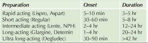
Fig. 23.1: Craniosynostosis: Common skull shapes.
multiple/severe sutural involvement may be associated with neuro-ocular complications and needs early craniotomy to prevent further restriction of brain growth.
Lacunar skull is characterized by bony defects in frontal/parietal regions of skull, which appear as irregular patches of rarefaction of X-rays. It is usually seen in craniosynostosis or associated with meningocele.
Microcephaly is an important cause of mental retardation, defined as head circumference lt; 3rd centile for age. Etiologically, it may be classified as primary or inherited defect and secondary, due to in utero or early postnatal insult to growing brain (see Table 18.25).
Clinically, microcephaly is associated with reduced velocity of postnatal head growth and developmental retardation, along with seizures and other features of chromosomal disorder or intrauterine infections. Abnormal facial appearance with slanting forehead and prominent nose and ears are more common in primary microcephaly (D/D secondary microcephaly)
Diagnosis of microcephaly per se rests on head circumference, though etiological diagnosis requires neuroimaging, karyotyping and serological studies for intrauterine infections. Salient differentiating features between microcephaly and craniosynostosis are given in Table 23.1.
Management of microcephaly is directed towards the primary cause, though developmental outcome is universally poor.
Facial dysmorphism is an important indicator of chromosomal disorders or underlying systemic disorders. Apart
TABLE 23.1: D/D microcephaly vs Craniosynostosis
AF: Anterior fontanel, MR: Mental retardation, *see Fig. 23.1
1 signs of chromosomal defects or intrauterine infections
2 except secondary neuro-ocular abnormalities
from abnormal facial appearance in microcephaly or craniosynostosis, discussed earlier, other important facial defects are as follows:
Hypertelorism, i.e. wide separation of eyes due to relative overgrowth of lesser wings of sphenoid, is confirmed by measuring interorbital distance. A Canthal index (inner canthal distance/outer canthal distance ? 100) of gt;38-40 at any age suggests hypertelorism.
Hypertelorism is seen in many chromosomal disorders, e.g. Down syndrome, craniosynostosis, e.g. Alport/Crouzon syndrome, and CNS anomalies, e.g. frontal encephalocele. Pseudohypertelorism is common in children with broad nasal bridge or epicanthic folds. Hypotelorism with Canthal index of lt;32, is relatively uncommon, usually associated with craniosynostosis, Patau syndrome (trisomy 13) and CNS defects, e.g. holoprosencephaly.
Depressed nasal bridge may be familial, chromosomal (mongolism), traumatic or post-infective, e.g. in syphilis. Micrognathia/retrognathia (Mandibular hypoplasia) is an important craniofacial abnormality in many syndromes and indicates defective development of first visceral/aortic arch in fetus.
Pierre Robin syndrome is characterized by a triad of: (a) micrognathia with relative macroglossia, (b) cleft palate, and (c) glossoptosis, with/without other defects, e.g. CHDs or mental retardation. Most cases die in infancy due to choking or aspiration, though survivors show good jaw growth with normalization of facial profile by ~5 years. These babies should be nursed in prone position with gavage feeding during early life to prevent choking. Temporary fixation of the tongue to alveolar margins may be necessary in severe cases to prevent glossoptosis.
Treacher-Collins syndrome is characterized by: (a) typical triangular face due to severe mandibular hypoplasia, (b) ocular malformations, e.g. antimongoloid slant or lid coloboma, and (c) ear deformities, e.g. microtia, absence of ear canal, preauricular skin tags or sinuses/dimples on a line from ear tragus to angle of mouth. Deafness, CHDs and mental retardation are common.
Prognathia (mandibular hyperplasia) may be familial or seen in chromosomal disorders or endocrinal defects, e.g. acromegaly.
23.2.2