DISORDERS OF PERITONEUM
Peritoneal disorders may be inflammatory (peritonitis) or non-inflammatory (ascites).
Acute peritonitis is either spontaneous or secondary (Table 14.26). While spontaneous peritonitis denotes peritoneal infection via hematogenous/lymphatic route from a source outside the abdomen, secondary peritonitis denotes extension of infection from an abdominal viscera.
Etiology: Spontaneous bacterial peritonitis (SBP) is usually caused by a single pathogen, e.g. S. pneumoniae,S. fecalis, Staph. aureus or gram negative organisms, e.g. E.coli and Kleb. pneumoniae. secondary peritonitis is usually polymicrobial.
TABLE 14.26: Causes of peritonitis
• Primary (spontaneous)
- Generalized septicemia
- Infected ascites, e.g. nephrotic syndrome, cirrhosis
• Secondary (extended infection or perforation)
- Intestines: Obstruction, appendicitis, diverticulitis
- Umbilical sepsis in newborns
- Liver: Hydatid cyst, pyemic/amebic abscess
- Biliary: Cholecystitis, worms, gallstones
- Acute pancreatitis
- Lymph node: Tuberculosis
- Foreign body: VP shunt
- Traumatic/post-surgical infection
SBP is an important complication of nephrotic syndrome, usually caused by capsulated organisms due to complement deficiency. On the other hand, peritonitis associated with ventriculoperitoneal shunt is usually caused by Staph. epidermidis, Staph. aureus and Candida albicans.
Clinically, acute bacterial peritonitis presents with high fever, diffuse abdominal pain and vomiting. On examination, these cases appear toxic with following typical features:
• Silent abdomen, i.e. restricted breathing movements to avoid pain,
• Rebound abdominal tenderness, guarding and/or boardlike rigidity,
• Diminished or absent bowel sounds.
Free fluid may be present and circulatory shock is common. Loss of liver dullness indicates pneumoperitoneum.
Presence of ascites in nephrotic syndrome or cirrhosis with pain or fever favors diagnosis of spontaneous peritonitis.Diagnosis must be suspected clinically, supported by polymorphonuclear leukocytosis and abdominal skiagram showing generalized haziness with sparse bowel gas shadows. Presence of free air under diaphragm (perforation) or multiple air-fluid levels (obstruction) indicate secondary peritonitis. USG with abdominocentesis is indicated in all cases to confirm the presence of fluid and differentiate ascites from peritonitis (proteins gt;2.5 gm/dl, cell count gt;250 cells/ mm3 with polymorphonuclear predominance), as well as for microbiological studies.
Management is largely conservative and includes:
• IV antibiotic therapy, which must be started immediately and empirically with Cefotaxime and an aminoglycoside, modified after culture reports. Additional IV metronidazole is recommended in secondary peritonitis. Therapy should be continued for 7-10 days.
• Supportive measures include no oral feeds, nasogastric decompression, fluid and electrolyte correction, parenteral nutrition and treatment of shock with volume expanders and vasopressors.
• Surgical intervention is needed only for underlying perforation or if peritoneal abscesses have developed.
Tubercular peritonitis, common in Indian children, results from spread of infection from regional lymph nodes and presents as a subacute peritonitis or ascites. Despite larger fluid collection, abdominal signs are less marked than in bacterial peritonitis and ascitic fluid is typically straw colored exudate with lymphocytic hypercellularity and cobweb formation on standing. USG/CT may reveal underlying abdominal lymphadenopathy or intestinal tuberculosis. Treatment includes specific anti-tubercular therapy with or without steroids (Ch 10.13, 14.12).
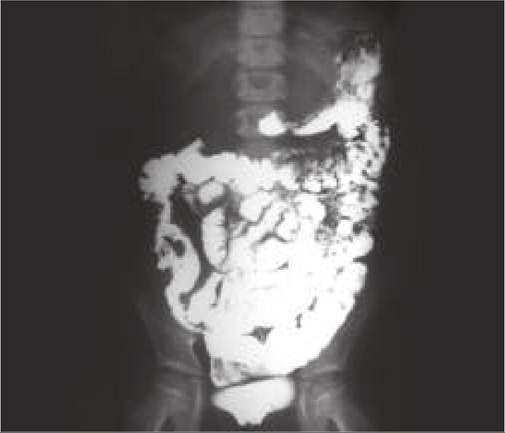
Fig. 14.21: Ascites.
Ascites denotes accumulation of any fluid within peritoneal cavity (Fig.
14.21). However, this term is generally reserved for serous and sterile fluid collections, while exudative ascites is referred as peritonitis.Etiology: Congenital malformations of thoracic duct or hydrops fetalis are common causes of ascites in newborns. In older children, it may be mechanical (altered hydrostatic:oncotic pressure balance), inflammatory or traumatic in origin (Table 14.27).
TABLE 14.27: Causes of ascites in children
Transudative ascites
• 4 Plasma oncotic pressure (hypoproteinemia)
- Hepatic: Cirrhosis, acute liver failure
- Renal: Nephrotic syndrome
- Nutritional: PEM
• Hydrostatic pressure
- Cardiac: CCF, pericardial effusion
- Vascular: Budd-Chiari syndrome
- Hepatic: Congenital hepatic fibrosis
- Portal hypertension
• Miscellaneous
- VP shunt
- Peritoneal dialysis
- Connective tissue disorders (Serositis)
Exudative ascites (peritonitis)
• Primary
- Tubercular peritonitis
- Spontaneous bacterial peritonitis
• Secondary
- Intestinal perforation
- Pancreatitis or ruptured pancreatic duct
- Biliary tree anomalies/injury (Biliary ascites)
- Urinary tract anomalies/injury (Urinary ascites) Hemorrhagic ascites
• Malignancies
• Traumatic (penetrating or blunt abdominal trauma)
• Splenic rupture
Chylous ascites
• Thoracic duct anomalies or injuries
Diagnosis: Abdominal distension is the hallmark of ascites, though other causes, e.g. gaseous distension, organomegaly or pot belly must be excluded. Depending on the amount of fluid collection, various clinical signs include:
• Flank fullness and Puddle sign (lt;50 ml fluid)
• Shifting dullness/horse-shoe dullness for moderate ascites, and
• Fluid thrill in large ascites.
Investigations: USG is the best tool to detect minimal ascites and for diagnostic paracentesis. Important characteristics of ascitic fluid include:
• Co lor: Watery (transudate), straw-colored (tubercular), hazy or pus (bacterial peritonitis), bloody (traumatic/ hemorrhagic), milky-white (chyle), greenish (biliary), tea-colored (pancreatitis).
• Proteins: Transudative (lt;2.5 gm/dl) or exudative (gt;2.5 gm/dl). As ascitic fluid hypoproteinemia is common in generalized hypoproteinemic states including SAM. A more informative test is to calculate serum: ascitic fluid albumin ratio. A ratio of gt;1.1:1 suggests portal hypertension.
• Leukocyte count of gt;500 cells/mm3 indicates exudative pathology, specially if gt;50% of them are polymorphs.
Other tests, e.g. bacterial and tubercular culture (peritonitis), triglycerides (chylous ascites), amylase (pancreatic etiology), LDH (spontaneous bacterial peritonitis) are also useful in diagnosis.
Management: Apart from treatment for primary cause, general management of ascites includes:
• Bed rest to decrease the discomfort as well as reduce renin-angiotensin activity, which is higher in upright position.
• Dietary restriction of sodium, though fluid restriction is not necessary unless serum sodium is lt;120 mEq/L.
• Diuretics, preferably potassium-sparing ones, e.g. spirolactone may be used in severe cases with caution to avoid hypovolemia.
• Therapeutic abdominocentesis carries the risk of infection in sterile ascites and recommended only in cases with marked discomfort and respiratory distress. Infected peritoneal fluid must be treated as peritonitis, discussed earlier.
BIBLIOGRAPHY
1. Srinivas S et al. Constipation in children standard treatment Guidelines. Indian Academy of Pediatrics. 2022.
2. Yachha S. Management of childhood functional constipation: Consensus practice guidelines of Indian Society of Pediatric Gastroenterology, Hepatology and Nutrition and Pediatric Gastroenterology Chapter of Indian Academy of Pediatrics. Indian Pediatr. 2018;55:885.
3. Basavaraja GV et al. Acute gastrointestinal bleed standard treatment guidelines. Indian Academy of Pediatrics. 2022.
4. Gupta R et al. Cleft lip and palate. Standard treatment guidelines. Indian Academy of Pediatrics. 2022.
5. Chokani R et al. Dental Caries.
Standard treatment guidelines. Indian Academy of Pediatrics. 2022.6. Indian Academy of Pediatrics: Oral Thrush. Standard Treatment Guidelines 2022.
7. Indian Academy of Pediatrics. Gastroesophageal Reflux Disease. Standard Treatment Guidelines 2022.
8. Mohan N et al. Diagnosis and management of gastroesophageal reflux disease in children: Recommendations of Pediatric Gastroenterology Chapter of Indian Academy of Pediatrics, Indian Society of Pediatric Gastroenterology, Hepatology and Nutrition (ISPGHAN) Indian Pediatr. 2021;58:1163.
9. Cherukuri N et al. Acute watery diarrhea. Standard treatment guidelines. Indian academy of Pediatrics. 2022.
10. Yachha S et al. Indian Academy of Pediatrics Consensus Guidelines for Probiotic Use in Childhood Diarrhea. Indian Pediatr. 2022;59:543.
11. Riyaz A et al. Persistent Diarrhea Standard treatment Guidelines. Indian Academy of Pediatrics. 2022.
12. Indian Academy of Pediatrics: Cow Milk Protein Allergy. Standard Treatment Guidelines 2022.
13. Matthai J et al. Guidelines on Diagnosis and Management of Cow's Milk Protein Allergy. Indian Pediatr. 2020;57:723.
14. Jaidev MD et al. Food Allergy. Standard treatment guidelines. Indian Academy of Pediatrics. 2022.
15. Kapoor A et al. Pediatric Inflammatory Bowel Disease. Indian Pediatr. 2016;53:993.
More on the topic DISORDERS OF PERITONEUM:
- Cirrhosis
- Ascites
- CLINICAL EVALUATION OF GIT DISEASE
- Agrawal M.. Textbook of Pediatrics. 3rd ed. — CBS Publishers,2025. — 973 p., 2025
- Hyposplenic states
- AMEBIASIS
- LIVER ABSCESS
- SPONTANEOUS SYMMETRY-BREAKING
- Chemically hazardous substances, ecotoxicology
- ABDOMINAL PAIN