INFANTILE HYPERTROPHIC PYLORIC STENOSIS
Infantile hypertrophic pyloric stenosis (IHPS) denotes narrowing of gastric outlet due to the unexplained thickening and hypertrophy of the pylorus muscle, primarily the circular layers.
Incidence: IHPS is the commonest cause of gastric outlet obstruction (~1-4/1000 live births), more common in males (2-5:1), first-born babies and offspring of the parents (specially mothers) with history of IHPS.
Etiology: In IHPS, pyloric hypertrophy is minimal at birth and develops gradually over first few weeks. Many genetic, familial and environmental factors have been attributed for this hypertrophy, e.g. (a) curdled milk, leading to mucosal edema and secondary pyloric hypertrophy, (b) less number and maturity of ganglion cells, (c) elevated gastrin levels and (d) deficiency of nitric oxide in pyloric muscle.
Clinically IHPS usually manifests from 2nd to 3rd week of neonatal life with:
• Recurrent projectile vomiting of curdled milk, after 20-30 minutes of feeding. Vomiting is essentially non-bilious, occasionally streaked with blood, and increases gradually in frequency, amount and force. After vomiting, baby is hungry and wants to be fed again.
• Failure to thrive, dehydration and constipation due to persistent vomiting. Infant is typically anxious, anemic and irritable/lethargic.
• Abdominal examination may reveal a firm, moveable, olive-shaped, supraumbilical lump on right side, which can be rolled between fingers. Lump is better palpable from the left side and immediately after feeding. Visible peristalses from left to right side are common after feeds, to overcome obstruction (Fig. 14.10A).
Fig. 14.10A: Infantile hypertrophic pyloric stenosis: Abdominal lump.
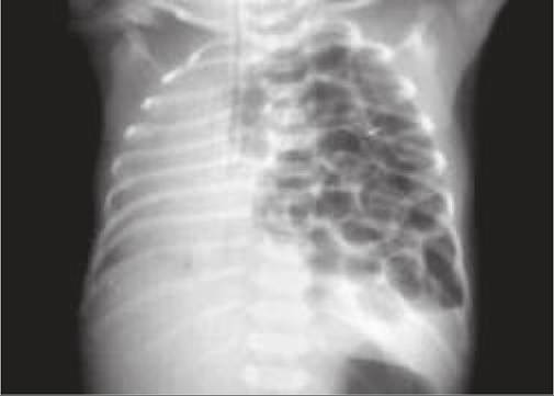
Fig.
14.10B: Infantile hypertrophic pyloric stenosis: Target (doughnut) sign on USG.• Obstructive jaundice may develop in ~2%, due to pressure of hypertrophied pylorus on ampula of Vater.
Associated anomalies like Down syndrome, Turner syndrome and tracheoesophageal fistula may be present. Diagnosis rests on typical appearance of pyloric lump, supported by:
• Barium meal showing—(i) long and narrow pyloric canal, (ii) string sign, i.e. passage of only a streak of barium, (iii) double-track sign due to crowding of mucosal folds in pyloric channel, and (iv) Shoulder sign due to bulging of pyloric muscles into antrum.
• Ultrasonography showing Target (Doughnut) sign due to hypertrophied ring of pyloric muscle around echogenic mucosa (Fig. 14.10B). Pyloric muscle thickness of gt;13 mm and length of pyloric channel gt;17 mm on USG is pathognomonic.
Treatment: While these cases need surgery as early as possible, most of them are too sick at the time of diagnosis with dehydration and electrolyte disturbances and need to be stabilized before surgery.
Pre-operative management includes—(a) nasogastric decompression, (b) correction of dehydration/electrolyte disturbances and (c) IV alimentation.
Hypochloremic metabolic alkalosis is commonest electrolyte abnormality in IHPS, due to loss of chlorides in vomiting. Normal saline or N/2 dextrose saline with 30-50 mEq/L of potassium is preferred for initial parenteral therapy with close monitoring of urine output and serum bicarbonate levels. Untreated alkalosis also increases the risk of post-operative apnea.
Ramstedt's pyloromyotomy is the surgery of choice in IHPS, in which hypertrophied pyloric muscles are split without cutting the mucosa and spread apart till mucosa prolapses into the pyloric incision. Oral feeding may be initiated after 12-24 hours, though some vomiting
TABLE 14.10: Causes of acute gastritis/ulcers
• Drugs: NSAIDs, iron, chloroquine, steroids
• Septicemia: Gm -ve infections, endotoxemia
• Dehydration/shock
• Severe hypoxia, e.g.
acute respiratory failure• Cushing ulcers - post-surgery, head injury
• Curling ulcers - burns
due to pyloric wall edema may continue for a few days. Laparoscopic pyloromyotomy or endoscopic balloon dilatation of pylorus are other options.
Other important gastric disorders are as follows:
Acute gastritis is usually caused by drugs, e.g. chloroquine or stress ulcers in critically sick children (Table 14.10) and presents with recurrent vomiting, epigastric pain and hematemesis in severe cases.
Treatment includes—(a) Antacids, (b) H2 receptor antagonists, e.g. Ranitidine (4-6 mg/kg/day q12hr), (c) Proton-pump inhibitors, e.g. omeprazole, and (d) mucosal- coating agents, e.g. sucralfate.
Chronic gastritis (peptic ulcer disease) in children has been strongly correlated with H. pylori infections, though family history of peptic disease is present in 25-50% cases. Zollinger-Ellison syndrome is a rare cause of multiple duodenal ulcers and diarrhea, due to gastrin-producing islet-cell tumor of pancreas.
Helicobacter pylori is a gram-negative, urease-producing organism, which colonizes ~50% children by 10 years of age and lives in the mucus overlaying gastric antrum. Infection is acquired by man-to-man transmission via feces, vomitus, saliva, etc. in overcrowded and unsanitary conditions. Although frequently associated with duodenal ulcers, whether the organism causes the ulcer or simply prevents its healing, is unclear.
Clinically, most H. pylori infections in childhood are asymptomatic, though some may present with recurrent abdominal pain.
Diagnosis is established by—(a) endoscopic mucosal biopsy from ulcer site using rapid urease test, or (b) breath test, to demonstrate urease production by increased excretion of radio-labeled CO2 after ingestion of C14/13 labelled urea.
Treatment is recommended only in symptomatic cases with triple-drug therapy for 2 weeks to eradicate infection. It includes an oral combination of clarithromycin (250500 mg) or amoxicillin (500-750 mg) with metronidazole (250-500 mg) and PPI, e.g.
omeprazole (20-30 mg), using lower range of doses mentioned against each drug for children 15-24 kg and higher range for those gt;25 kg.Bezoars, i.e. an accumulation of exogenous matter in stomach or intestine, are more common in mentally challenged children and in females. Depending on type of matter, bezoars are called trichobezoars (hair), phytobezoar
(plant/animal material), Iactobezoar (casein/calcium of top milk), etc.
These cases usually present in second decade with recurrent abdominal pain, vomiting, palpable lump in left hypochondrium and sub-acute obstruction. Failure to thrive is common.
Abdominal X-ray may show bezoars in plain or barium-contrast films (Fig. 14.11), though endoscopy is useful for diagnosis as well as removal. Lactobezoars usually resolve spontaneously after 24-48 hours.
14.9
More on the topic INFANTILE HYPERTROPHIC PYLORIC STENOSIS:
- Stenosis
- Mitral Stenosis
- Agrawal M.. Textbook of Pediatrics. 3rd ed. — CBS Publishers,2025. — 973 p., 2025