SMALL BOWEL MALFORMATIONS
Small bowel malformations are leading causes of intestinal obstruction in newborns and young infants, though many of them present in later life or remain asymptomatic throughout the life.
Duodenal atresia: Duodenal atresia is rare (~1:10,000 live births) but important defect, characterised by developmental failure of the recanalization of duodenal lumen after solid phase of intestinal development, producing stenosis, atresia or a mucosal web in the duodenal lumen, usually at the site of papilla of vater.
Chromosomal abnormalities, e.g. Down syndrome are present in ~25% cases, while gt;30% have other gut malformations, e.g. malrotation, esophageal atresia, anorectal anomalies, etc. or congenital heart diseases. Half of these cases are preterms. Polyhydramnios is present in gt;50% cases, due to failure of the absorption of amniotic fluid.
Clinically, these cases present within 24 hours of birth with—(a) bilious vomiting/gastric aspirate without abdominal distension, and (b) visible peristaltic waves in upper abdomen. Jaundice is present in 1/ 3rd cases due to blockade of ampula of Vater.
Diagnosis rests on X-ray abdomen showing pathognomonic double-bubble sign, i.e. one air-fluid level in the distended stomach and another in the first part of distended duodenum. Rest of the gut has no air shadows (Fig. 14.12A).
Prenatal diagnosis is possible on USG, while postnatal USG is necessary before surgery to exclude other malformations or causes of duodenal obstruction, e.g. annular pancreas, peritoneal bands, volvulus, etc.
Management: Immediate duodenoduodenostomy or duodenojejunostomy is necessary in all cases, after preoperative stabilization with nasogastric decompensation, fluid/electrolyte correction, etc. and exclusion of other malformations. Postoperative oral feeds may be started after ~1 week, after disappearance of duodenal ileus.
Jejunoileal atresia: Jejunoileal atresia is twice more common than duodenal atresia and have been attributed to intrauterine infections or vascular accidents.
These defects are of four types—type I, with an intraluminal diaphragm; type II, with a solid cord between proximal and distal segments; type III, with blind loops on both ends of bowel with mesenteric defects; and type IV, with multiple atretic segments. Type III is most common (35%).
Clinically, these defects invariably present within 24-48 hours with meconium ileus and acute intestinal
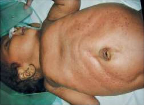
Fig. 14.12B: Jejuno-ileal atresia (Triple bubble sign).
obstruction. Polyhydramnios is present in ~ 25% cases. Diagnosis rests on plain X-ray abdomen, showing three fluid levels (Triple-bubble sign, Fig. 14.12B), meconium ileus (ground-glass appearance in right lower quadrant) and/or evidence of perforation, i.e. gas under diaphragm. Presence of peritoneal calcifications suggests meconium peritonitis.
Treatment involves immediate surgical exploration with resection-anastomosis or proximal jejuno/ileostomy, after pre-operative stabilization.
Malrotation (Volvulus): Most of the mid-gut in early fetal life lies outside the abdomen cavity, but re-enters later with a rotation around the axis of superior mesenteric artery, which allows placement and fixation of intestinal structures at normal locations. Malrotation or non-rotation leads to abnormal location of these structures, commonest defect being the failure of the cecum to move in right lower quadrant.
Volvulus refers to an acute or recurrent symptomatic complication of malrotation, which need immediate intervention.
Clinically, most cases of malrotation (60-80%) present in infancy as volvulus with acute intestinal obstruction, while rest may present as chronic obstruction with recurrent abdominal pain and vomiting. However, ~25-50% cases with mild defects remain asymptomatic or present beyond childhood.
Diagnosis usually depends on USG or barium contrast studies. Plain X-rays are often non-specific
Treatment: Surgical intervention is indicated in all diagnosed cases irrespective of age, usually along with incidental appendectomy.
Meckel's diverticulum: Meckel's diverticulum is a
3-6 cm out-pouch of the ileum along the anti-mesentric border, ~50-75 cm away from ileocecal junction. It represents attenuated omphalomesenteric duct, which connects the yolk sac with developing embryo before development of placenta.
Pathologically, it is a true diverticulum with all intestinal layers, lined by the heterotopic gastric mucosa. Pancreatic tissue or jejunal/colonic mucosa may also be present.
Incidence: Meckel's diverticulum is the commonest congenital malformation of gut, present in 1.5-2.0% of normal population, more common in males (2:1). It may also be associated with other gut malformations, e.g. esophageal atresia, anorectal anomalies, etc. or Crohn disease.
Clinically most cases are asymptomatic. Symptomatic cases usually present in first two years of life with complications, e.g. (a) intermittent painless rectal bleeding, (b) acute intussusception with diverticula as its lead point, or (c) acute abdominal pain due to diverticulitis or impacted roundworms in diverticula. Rarely, tumors, e.g. carcinoid tumor may originate from diverticula in late life.
Diagnosis: A radionucleotide scan with Tc99m to delineate gastric mucosa is the best diagnostic method to detect Meckel's diverticulum with gt;85% sensitivity and specificity (Meckel's scan). Sensitivity may be further enhanced with prior administration of cimetidine, pentagastrin or glucagon.
Other investigations, e.g. rectosigmoidoscopy may be necessary to exclude other causes of rectal bleeding.
Management: Open or laparoscopic wedge resection of the diverticulum or segmental resection of the ileum bearing the diverticulum is indicated in all symptomatic cases after pre-operative stabilization. In asymptomatic, incidentally detected diverticula, resection is advisable in cases with—(a) diverticular length gt;2 cm, (b) younger age lt;40 years and (c) presence of heterotopic mucosa.
Peutz-Jeghers syndrome is a rare autosomal dominant disorder, characterized by multiple gastric and intestinal polyps with perioral and oral hyperpigmentation and presenting with recurrent abdominal cramps, intussusception and occasional gastrointestinal bleeding.
14.9.2
More on the topic SMALL BOWEL MALFORMATIONS:
- Inflammatory Bowel Disease
- Agrawal M.. Textbook of Pediatrics. 3rd ed. — CBS Publishers,2025. — 973 p., 2025
- CONGENITAL LUNG MALFORMATIONS
- 12 Congenital Anomalies
- Screening for chromosomal abnormalities
- Chapter 30 Pelvic Support Defects, Urinary Incontinence, and Urinary Tract Infection
- 32 Colonic vascular ectasia in a dog
- Management of women with uncertain early pregnancy diagnosis
- Haemorrhagic gastroenteritis in a dog
- Anorectal physiology and function