32 Colonic vascular ectasia in a dog
Initial presentation
Collapse and haematochezia
Signalment: 5-year-old neutered male Bichon Frise, body weight 8.5 kg
Case history
The dog had presented to the referring veterinary surgeon 1 month previously with a history of having collapsed.
Clinical examination at that time had been unremarkable. The only abnormal finding was highly regenerative anaemia, with a packed cell volume (PCV) of 0.22 l/l (reference range 0.37-0.55 l/l) and a reticulocyte count of 335 ? 109∕l (reference range >60 ? 109∕l, indicates a regenerative anaemia). Empiric treatment had been prednisolone and ferrous sulphate (doses not known).Since then the dog had been increasingly lethargic and his appetite had been poor. His faeces were of normal consistency, but frequently were dark in colour and also contained bright red blood (haematochezia) and some mucus. The owners reported that he had lost about 0.5 kg during this time.
He was de-wormed every 6 months and his vaccinations were current. His regular diet was a commercial dry dog food.
Physical examination
The dog was responsive but lethargic and quiet. His body condition score was 4/9 with evidence of recent weight loss. His mucous membranes were very pale and slightly dry. His capillary refill time was 1%). His platelet count was within the reference range, as were coagulation parameters (activated partial thromboplastin time and prothrombin time). Serum chemistry results were within the reference range other than a slightly decreased albumin of 23.6 g/l (reference range 26-35 g/l) which may have been due to an intestinal disorder causing malabsorption or from the blood loss.
After transfusion his PCV increased to 0.27 l/l and the murmur disappeared. His heart rate deceased to 60 bpm, although an ECG showed no abnormalities other than a wandering pacemaker (not considered to be pathological).
Faecal samples were negative for parasites and enteropathogenic bacteria, but highly positive for faecal occult blood.
Imaging
Radiography and abdominal ultrasonography showed no abnormalities other than food present in the stomach after 12 hours of fasting, indicative of delayed gastric emptying.
Endoscopy
On endoscopy, the gastric and duodenal mucosa appeared grossly normal. The colon had numerous areas of raised, tortuous, dilated blood vessels which were diffusely spread throughout the length of the colon (Fig 32.1). Histology of the mucosal sample from the duodenum was consistent with mild, chronic duodenitis. Histology of the sample from the colon was interpreted as moderate chronic active colitis.
Fig 32.1
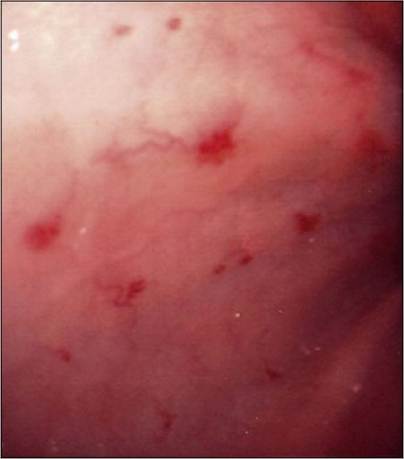
Endoscopic view of colon showing abnormal vessels
Diagnosis
The severity of the dog’s signs and the severity of the gross appearance of the colon, along with the lack of response to corticosteroids, were not consistent with inflammatory bowel disease. A diagnosis of colonic vascular ectasia was made from the characteristic gross morphologic abnormalities visualized during colonoscopy.
Treatment and outcome
Palliative therapy with tylosin in addition to the prednisolone was initially tried in this case, but with no success. As the lesions were diffusely distributed throughout the colon, laser coagulation or partial colonic resection were not feasible options. A subtotal colectomy was discussed with the owners. This surgery in dogs is not as successful as it is in cats with dilated idiopathic megacolon as reservoir faecal incontinence is a common complication in dogs. Further medical management with hormone therapy was discussed; however, the dog had another severe bleeding episode and unfortunately the owners chose to have him undergo euthanasia.
Discussion
Colonic vascular ectasia, also known as angiodysplasia, is the presence of dilated arteries, veins and lymphatic vessels lined with a single layer of endothelial cells within the mucosa and submucosa of the colon.
Haemorrhage is caused by disruption of the blood vessels, which often results in chronic haematochezia and an iron deficiency anaemia characterized by hypochromia and microcytosis. Melaena may also be present if the lesions extend into the small intestine or if the transit time in the colon is slow enough to allow digestion of the haemoglobin.The aetiology is unknown, although theoretically colonic distension from chronic constipation could lead to submucosal venous occlusion and dilation. However, it has not been reported in cats with acquired megacolon due to obstipation and neither this case nor most of the reported cases had a history of constipation or tenesmus.
Diagnosis of the disorder is based upon assessment of the colonic mucosa at endoscopy and the characteristic appearance. The lesions are not usually visible from the serosal surface and would be missed at exploratory laparotomy.
Subtotal colectomy and partial colectomy (aided by intra-operative endoscopy to locate the lesions in one case) have been used successfully to treat the disorder.
Successful use of oral oestrogen-progesterone therapy has been used in humans with the condition and has been reported in two canine cases. In humans, this therapy does not lead to regression of the lesions, but it was associated with a reduction in the number of blood transfusions necessary to maintain stable to normal haemoglobin concentrations. The mechanism of action of the drug combination is not well understood, but theories include an induction of squamous metaplasia, restoration of the continuity of the endothelium of abnormal vessels, primary effects on blood coagulation and stasis of blood in the mesenteric microcirculation. In reported canine cases, while the lesions persisted as in humans, the haematochezia resolved and no further transfusions were necessary. The dose used was 2 μg∕kg ethinyl oestradiol po q 24 hours and 23 μg/ kg of norethindrone acetate po q 24 hours. Ferrous sulphate at 18 mg∕ kg po q 24 hours was also administered.
The potential risk of the oestrogen-progesterone therapy is bone marrow toxicity resulting in pancytopenia, as well as alopecia, cystic endometrial hyperplasia and pyometra, although the doses used in these two dogs appeared to be low enough to avoid these side effects.
Prognosis
There have been insufficient cases reported to predict the prognosis for these cases. Surgical management for focal lesions has been reported to be successful and in limited numbers, medical management has also been successful for cases in which surgery was not possible. It is likely that many of these cases die or are euthanized without a diagnosis.