Screening for chromosomal abnormalities
The most common chromosomal defects are trisomy 21 (Down syndrome), accounting for 50% of the chromosomal abnormalities; 25% are trisomy 13 (Patau syndrome) or trisomy 18 (Edward syndrome); 10% are monosomy X (Turner syndrome); 5% are triploidies; and 10% are other aneuploidies.
First-trimester screening
The first-t rimester ultrasound scan was initially introduced with the only purpose of measuring the crown-rump length (CRL) and dating the pregnancy accordingly.
After the initial screening tests based on maternal age alone and maternal serum biochemistry on the second trimester (15, 16), the screening programmes moved towards the first trimester for the purpose of screening for chromosomal abnormalities. This started with the introduction of the NT as the main method of screening combined with the biochemical markers (17, 18). The NT is the measurement of the ultrasound echo-free space (translucency) between the skin and the soft tissue overlying the cervical spine. It needs to be performed between 11 weeks and 13 weeks and 6 days (or a CRL between 45 and 84 mm), on a sagittal section of the fetal head and with appropriate magnification of the image (Figure 11.1).
While early studies used fixed cut-off values for NT, it is now understood that NT is a dynamic measurement that increases as gestational age advances. The 95th centile is therefore dependent on the gestational age (and, therefore, CRL). NT is increased (>95th centile) in approximately 70% of fetuses with trisomy 21. An increased NT can also be associated with other chromosomal abnormalities (trisomy 13, trisomy 18, Turner syndrome, and triploidy among others), genetic conditions, and fetal structural abnormalities (most commonly cardiac defects) (19, 20). It is also known that the higher the value is, the higher the risk of fetal death.
The NT is combined with the maternal serum biochemical markers (free beta-hCG and PAPP-A) to improve the detection rate.
Typically, in trisomy 21 the maternal serum free beta-hCG is increased to about twice as high and the PAPP-A is halved compared to euploid pregnancies (2, 21). In the case of trisomies 18 and 13, both beta-hCG and PAPP-A are decreased (22).Other sonographic markers described in the first trimester are the nasal bone (23), ductus venosus flow (24), and tricuspid regurgitation (25). The combination of maternal age, NT thickness, biochemical markers, and these three additional markers shows detection rates as high as 96% for trisomy 21 with a false-positive rate of 3% (26).
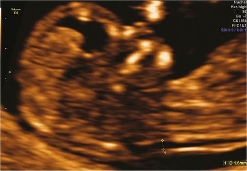
Figure 11.1 Mid-sagittal ultrasound view of the fetal head with measurement of the nuchal translucency at 12 weeks.
A detailed first- trimester scan performed by an experienced operator can detect a number of major structural abnormalities.
Trisomy 18 can be suspected in the first trimester in the presence of a strawberry-shaped head, heart defects, diaphragmatic hernia, oesophageal atresia, exomphalos, single umbilical artery, megacystis, radial aplasia, overlapping fingers, and talipes. Common ultrasound features of trisomy 13 that can be detected in the first trimester are holoprosencephaly, facial abnormalities, cardiac abnormalities, echogenic kidneys, exomphalos, and postaxial polydactyly.
Second-trimester screening
Routine second-trimester ultrasound scanning was introduced in the United Kingdom in the 1980s. However, uniform criteria on how to perform and what to look for in this scan were only introduced recently by the NHS FASP.
During the first trimester, the most frequent marker of chromosomal abnormality is the increased NT. In the second trimester, each chromosomal defect can have its own specific structural abnormalities:
• Strawberry-shaped head: this feature is characteristic of trisomy 18, and is present in up to 80% of cases. The frontal skull is narrow with a flat occiput.
• Holoprosencephaly: the incidence at birth is 1:10,000. It includes a heterogeneous group of cerebral malformations as a result of a failed or incomplete cleavage of the forebrain. Around 30% of them are associated with chromosomal defects, in particular trisomy 18 and trisomy13, the latter sometimes associated with microcephaly. The holoprosencephaly can be associated with facial clefts or other defects and the incidence of chromosomal defects is higher if it is associated with extrafacial abnormalities.
• Choroid plexus cysts (CPCs): the presence of CPCs together with other structural abnormalities raises the suspicion of trisomy 18. However, isolated CPCs can be found in 2% of the fetuses in the second trimester and is mostly benign. They typically disappear by 28 weeks.
• Agenesis of the corpus callosum: the incidence is 1:1000 births and it is related to chromosomal defects, especially trisomy 18 and trisomy 13. It can be suspected by the absence of cavum septum pellucidum (CSP) in the biparietal diameter view or by the presence of abnormalities in the lateral ventricles.
• Ventriculomegaly: this is defined by the increase in the transverse diameter of the atrium of the lateral ventricle by more than 10 mm. It can be secondary to infection, haemorrhage, or obstruction or associated with genetic conditions or chromosomal defects in around 10% of cases. The most frequent are trisomies 21, 13, and 18 and triploidy. The incidence of chromosomal defects is higher with mild to moderate, rather than severe ventriculomegaly.
• Dandy-Walker malformation: there is a spectrum of defects of the cerebellar vermis, cystic dilatation of the fourth ventricle, and enlargement of the cisterna magna. The incidence of chromosomal defects in the presence of Dandy-Walker malformation is as high as 40%, more frequent in trisomy 18, trisomy 13, and triploidy.
• Facial cleft: the term facial cleft includes cleft lip and palate that may present together. The incidence of chromosomal defects is around 10%, most frequently trisomy 13 and trisomy 18.
• Micrognathia: this entity is common in numerous genetic syndromes. Among the chromosomal defects, trisomy 18 and trip- loidy are the most likely ones.
• Nasal bone hypoplasia: the presence of a small nasal bone on the sagittal view of the face is defined as a hypoplasic nasal bone and is seen in 60% of the fetuses with trisomy 21. Only 1-2% of normal fetuses present with this feature and it is considered the most sensitive and specific isolated second-trimester marker of trisomy 21 (27).
• Nuchal oedema: this is the equivalent of the increased NT thickness in the second trimester. It is defined as an increase in the subcutaneous tissue greater than 5 mm measured on a suboccipitobregmatic view of the head. It is present in about 30% of fetuses with trisomy 21. It can also be associated with infection and other genetic conditions.
• Cystic hygroma: this is a congenital malformation of the lymphatic system. On the ultrasound scan, it is possible to identify a bilateral swelling of the posterolateral aspect of the fetal neck. It is typically septated and often there is a thick midline septum corresponding to the nuchal ligament. It is strongly associated with chromosomal defects, mostly Turner syndrome but also trisomy 21 and trisomy 18. About 20% of the fetuses have a normal karyotype. There is also an association with generalized hydrops that carries a worse prognosis.
• Cardiac abnormalities: the presence of cardiac defects is seen in 90% of the fetuses with trisomy 13 and trisomy 18, 50% with trisomy 21, and 40% with Turner syndrome. Some studies have reported a 25% incidence ofchromosomal defects in the presence of a cardiac abnormality. The presence of intracardiac hyperechogenic foci is considered one of the soft markers of chromosomal defects on the second-trimester scan and may be seen in up to 25% of fetuses with trisomy 21 (27).
• Congenital diaphragmatic hernia: the incidence is around 1:4000 live births. It can be an isolated finding but in 30-50% of the cases there is an underlying chromosomal defect, most frequently trisomy 18 and tetrasomy 12p (Pallister-Killian syndrome).
• Oesophageal atresia: oesophageal atresia is a relatively common gastrointestinal tract defect with an incidence of 1:4000 births. It is associated with aneuploidy in 5% of the cases, most frequently trisomy 13 and trisomy 18. It can also be part of the VACTERL syndrome (vertebral abnormalities, anal atresia, cardiac defect, tracheo-oesophageal fistula, renal and radial limb abnormalities) or CHARGE (choanal atresia, renal abnormalities, tracheo-oesophageal fistula, micrognathia, cleft lip/palate, and exomphalos). The diagnosis is suspected if there is failure to visualize the stomach bubble or a very small stomach is seen on serial ultrasound scans. At later stages in pregnancy, it can be associated with polyhydramnios. Most cases are associated with a tracheo- oesophageal fistula, and antenatal identification is possible in less than half of all cases.
• Duodenal atresia: the incidence is 1:10,000 births. Up to 50% of the cases are associated with other abnormalities: cardiac malformations (10-20%), trisomy 21 (30%), skeletal dysplasias, and intrauterine growth restriction. The classical sign is the ‘double bubble' as a result of the enlarged stomach and proximal duodenum.
• Exomphalos: the reported incidence varies between 1:4000 and 1:7000 livebirths. In 40% of the cases there is an underlying
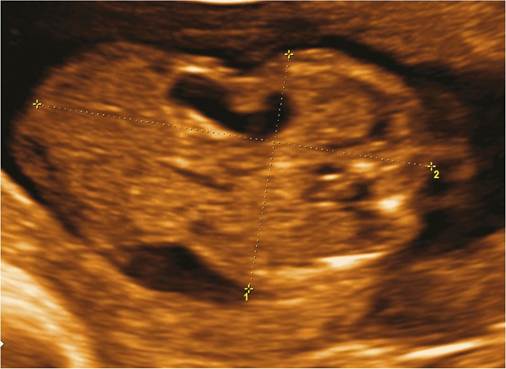
Figure 11.2 Exomphalos containing bowel and stomach.
chromosomal defect, usually trisomy 13 or 18, most commonly if there is only small exomphalos containing bowel rather than a large exomphalos containing liver as well as small bowel. Even in the absence of aneuploidy, additional structural abnormalities are encountered in 80% of the cases, cardiac defects in 30% of these. Other associations are Beckwith-Wiedemann syndrome, cloacal exstrophy, pentalogy of Cantrell, and Meckel-Gruber syndrome (Figure 11.2).
• Hyperechogenic bowel: this is found in 1-2% of all fetuses in the second trimester.
In 50% of the cases there is spontaneous resolution. It can be associated with cystic fibrosis, chromosomal abnormalities (in particular, trisomies 21, 18, or 13 and triploidy), intra-amniotic bleeding, congenital infection, growth restriction, and bowel obstruction.• Renal/urinary tract anomalies: although mild renal pelvic dilatation is relatively common, the presence of a mild hydronephrosis is also common in fetuses with trisomy 21 (10-25%). A diagnosis of moderate to severe hydronephrosis, polycystic kidneys, or renal agenesis is more common in trisomies 18 and 13. Megacystis (>7 mm longitudinal diameter in the first trimester) raises the possibility of trisomy 18.
• Skeletal dysplasias: there is a wide variety of skeletal dysplasias and the suspicion of any abnormality of the bones should trigger further investigations and detailed ultrasound examination to exclude other structural abnormalities. A short femur is present in 40% of the fetuses with trisomy 21; also characteristic are clinodactyly and the sandal gap sign. The presence of polydactyly, overlapping fingers, and rocker-bottom feet are classical signs of trisomy 18.
• Growth restriction: this is a common finding in many chromosomal defects and genetic syndromes, but not trisomy 21. Trisomy 18 and triploidy are classically associated with severe growth restriction from very early stages of pregnancy. In addition, in trip- loidy there is a marked disproportion of the head size compared to a marked reduction in the growth of the abdomen and femur.
In addition to a detailed second-trimester ultrasound scan, risk assessment for chromosomal abnormalities can be performed using maternal serum biochemistry (quadruple test). It combines maternal age with maternal serum levels of alpha-fetoprotein, free beta-hCG, unconjugated oestriol, and inhibin A and has a detection rate of 70-75% with a 5% false-positive rate (15, 16). Due to the low detection rate compared to the combined first-trimester screening, the use of this test is mainly limited to women who missed the first- trimester risk assessment using the combined test.
More on the topic Screening for chromosomal abnormalities:
- Screening for chromosomal abnormalities
- Prenatal diagnosis
- Universal screening and case finding
- Counselling for couples with genetic abnormalities and inheritable conditions
- REFERENCES
- 4 Preconception Counseling and Prenatal Care
- 12 Congenital Anomalies
- Invasive Prenatal Diagnosis in Multiple Pregnancy
- Structural abnormalities
- Screening for Fetal Abnormality in Multiple Pregnancy