Abnormal cytologic findings
Nuclear changes
Brick inclusions
These large blue crystalline rectangular inclusions are commonly noted in hepatocytes and are more common in older dogs (Figures 9.3, 9.8).
Brick inclusions have no known pathologic significance, do not contain heavy metals, and are acid-fast negative, in contrast to lead inclusions, which are positive with acid-fast staining (Richter et al., 1965; Reimer et al., 1973). 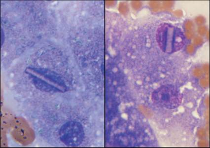
Figure 9.8 Brick inclusions are occasionally seen in hepatocyte nuclei; they have no known significance (Wright–Giemsa, 1,000? magnification).
Intranuclear cytoplasmic inclusions
Intranuclear cytoplasmic inclusions are, as the name implies, blebs of cytoplasm that appear to be within the nucleus (Figure 9.9). They are usually round, can vary in size, and are commonly thought of as a sample preparation artifact. They can be found in many cell types, including hepatocytes.
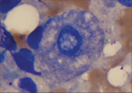
Figure 9.9 A hepatocyte with an intranuclear cytoplasmic inclusion is noted in this image. This finding is of unknown significance. The cytoplasm is also rarefied, consistent with glycogen or hydropic degeneration (Wright–Giemsa, 1,000? magnification). (Same animal as Figure 9.12.)
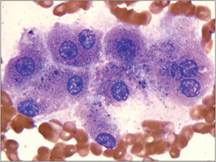
Figure 9.10 Many binucleate hepatocytes are found. This patient also had a mild suppurative and histiocytic inflammation (not shown) (Wright–Giemsa, 1,000? magnification).
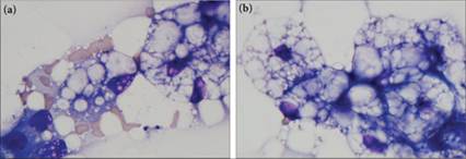
Figures 9.11a,b Domestic shorthair cat with lipid-laden hepatocytes.
(a) Note the morphologically more normal hepatocytes on the lower left of the image, progressing to cells filled with distinct lipid vacuolation in the upper right corner of the image. The origin of the highly vacuolated cells would be challenging to determine if viewed alone. (b) This cluster of hepatocytes is heavily lipid-laden and can be confused with adipocytes. Abundant free lipid is also noted as clear defects in the normally lightly blue extracellular fluid background of the images (Wright–Giemsa, 1,000? magnification).
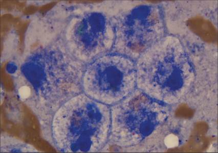
Figure 9.12 Hepatic FNA from a 16-year-old Yorkshire Terrier. Note how the cytoplasm of these hepatocytes is rarefied by foamy cytoplasmic clearing, consistent with glycogen or hydropic degeneration. Cytologically, this type of change contrasts with the distinct lipid vacuolation seen in Figures 9.11a, b (Wright–Giemsa, 1,000? magnification).
Intranuclear lead inclusions are observed infrequently, even after intentional dosing of dogs with lead (Hamir et al., 1983). On electron microscopy (EM) evaluation, these acid-fast inclusions are dense with irregular borders and are surrounded by a clear zone (Hegazy & Fouad, 2014).
Binucleation
Some binucleation is seen on most, if not all, canine liver aspirates. Rarely, trinucleate cells can be observed. Increased amounts of bi- and trinucleation are most commonly associated with increased mitotic activity, as occurs with a regenerative effort (Figure 9.10). In one study, the number of binucleate and trinucleate hepatocytes in apparently healthy patients was correlated with age; this may be associated with an increased incidence of nodular regeneration in older individuals (Stockhaus et al., 2002).
Anisokaryosis
Often concurrent with binucleation, mild to moderate anisokaryosis, or variably sized nuclei, will be noted in mitotically active hepatocytes. Additionally, benign and malignant hepatic and biliary tumors can display cellular atypia, including anisokaryosis. Therefore, although nuclear size changes are more commonly found cytologically in regenerative lesions, they are not pathognomonic for hepatocellular regeneration.
Cytoplasmic changes
As a consequence of their multiple roles and varied metabolic processes, atypical hepatocytes can accumulate a wide variety of cytoplasmic inclusions, including pigments and vacuoles. Routine cytologic preparations are not the most sensitive at distinguishing these inclusions, but additional staining procedures can be used to completely identify cytoplasmic inclusions when a definitive diagnosis is required. There are several cytoplasmic changes easily noted with Romanowsky stains, as described below.