Normal cytologic findings
Hepatocytes
Cytologically, hepatocytes are generally found in cohesive clusters and are the predominant cell type other than red blood cells (RBCs) in a liver aspirate. They are moderately sized, polygonal cells with abundant amounts of basophilic to pink granular cytoplasm, a centrally located round nucleus with finely stippled chromatin, and a single prominent nucleolus (Figures 9.3, 9.4a).
In health, there is typically minimal variability between hepatocytes. In the diseased liver, there can be hepatocytes with minimal cytologic changes juxtaposed with hepatocytes with marked pathologic changes. 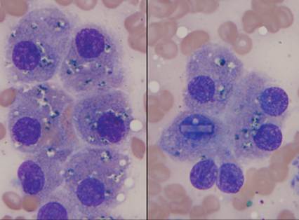
Figure 9.3 These polygonal cells are characteristic of hepatocytes. They have basophilic to pink granular cytoplasm, a centrally located nucleus with finely stippled chromatin, and a single nucleolus. Minimal pigment consistent with lipofuscin or other pigment (blue–black granular cytoplasmic material. An intranuclear brick inclusion is present (right panel) (Wright–Giemsa, 1,000? magnification).
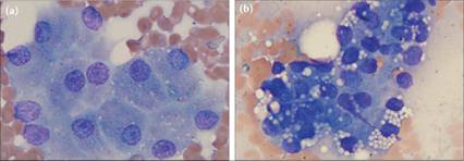
Figures 9.4a,b This canine liver aspirate has two populations of normal cells present. (a) The predominant cell population is morphologically normal hepatocytes. (b) There are also two clusters of biliary epithelial cells, which, compared with the hepatocytes, have less cytoplasm and distinct clear vacuolation (Wright–Giemsa, 1,000? magnification).
Biliary epithelial cells
Biliary epithelium is usually encountered in smaller amounts as cohesive epithelium in tubular and papillary formations (Figure 9.4b). In a review of healthy dogs, an average of three biliary cells per two slides was observed (Stockhaus et al., 2002).
However, this study also found that thicker samples had relatively more biliary epithelium present. Biliary epithelium of smaller bile ducts are low columnar to cuboidal cells with minimal to moderate amounts of cytoplasm, which is lightly to deeply basophilic and filled with distinct clear vacuoles and which are smaller than hepatocytes. The nuclei are commonly basally located and more condensed. Biliary epithelium from larger ducts is columnar and can be ciliated (Masserdotti, 2020).Other cells
Few small lymphocytes, neutrophils, mature adipocytes, and mast cells are encountered during evaluation of a healthy liver. Each of these cell types makes up less than 1% of cells counted in one canine study (Stockhaus et al., 2002). Mast cells can be found in higher numbers in normal canine hepatic aspirates when stained with Toluidine blue compared with May–Grünwald–Giemsa; however, even with better detection, they still total less than 1% of nucleated cells (Masserdotti, 2013). Additionally, samples of a healthy liver will typically have less than 0.1% of resident pigment-laden macrophages (Kupffer cells), mesothelium, eosinophils, and fibrocytes. When mesothelial cells are found, they will frequently be round individualized cells with basophilic to pink cytoplasm and prominent cytoplasmic blebs or will be present in cohesive sheets which have a cobblestone appearance (Figures 9.5–9.7). Due to its highly vascularized nature, hepatic aspirates often contain moderate to abundant amounts of peripheral blood elements, including RBCs, leukocytes, and platelet clumps. If peripheral blood elements are present solely as a result of the procedure, the relative numbers of cells will be similar to peripheral blood.
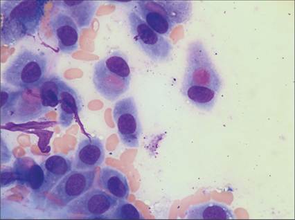
Figure 9.5 Five-year-old, male Coton de Tolear. Note the cohesive sheet of mesothelium in the lower left corner; these cells have a characteristic cobblestone appearance.
The centrally located, pink cytoplasmic material noted in several of the mesothelial cells can occasionally be observed. (Courtesy Dr. Francesco Cian.)
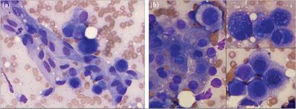
Figures 9.6a,b Liver aspirate from an Australian Shepherd Dog that had metastatic mammary carcinoma. (a) Sheets of normal mesothelium are present as an incidental finding. Note how these thin sheets can easily fold and wrinkle during slide preparation (Wright–Giemsa, 500? magnification). (b) Multiple neoplastic cells are also present (right panels), including a single macrocytic neoplastic cell with high N:C ratio (left panel), when compared with normal hepatocytes. Prominent cell–cell junctions (lower right panel) and multiple criteria of malignancy, including anisokaryosis, anisocytosis, anisonucleoliosis, and variable N:C ratio, are noted in the neoplastic population (all panels) (Wright–Giemsa, 1,000? magnification).
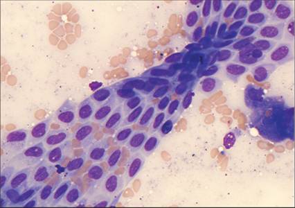
Figure 9.7 Note the distinct cobblestone appearance of the single layer of mesothelial cells that has folded over on itself (Wright–Giemsa, 500? magnification). (Same dog as in Figure 9.20.)