Function and organ architecture
The liver plays a vital role in many catabolic and anabolic processes. Individual hepatocytes can be viewed as factory workers standing at a blood-based conveyor belt, busily picking up components and altering them either for further manufacturing work at distant sites (other body systems) or disposal (excretion, usually through the biliary or urinary system).
As a result of this role, the liver has developed as a highly vascularized system with hepatocytes arranged as plates of cells, two layers thick, sandwiched between a fenestrated vascular capillary network (Figures 9.2a, b). Located within the hepatocyte bilayer is an interconnecting biliary network of fine canaliculi combining into large epithelium-lined ducts that lead to the gallbladder. This biliary network serves both as a pathway for excreta and as a method for the modification and recycling of metabolites. 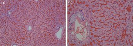
Figures 9.2a,b Histologic section of a canine liver. (a) The central vein is in the center of the image with plates of hepatocytes radiating to the portal triads along the periphery of the image. The hepatic sinusoids in this sample are filled with red blood cells (H&E, 100? magnification) (b) A closer image provides better detail of the hepatocytes and contents of the portal triad. Note the columnar epithelial cells within the bile duct, the hepatic artery surrounded by smooth muscle, and the large and open hepatic vein along the left of the image. A central vein filled with red blood cells is visible in the upper right corner of the image (H&E, 200? magnification).