Acidophilic Macrophage Pneumonia/ Epithelial Hyalinosis
Acidophilic macrophage pneumonia (AMP) is characterized by focal to diffuse accumulation of acidophilic crystals within macrophages, alveolar spaces, and airways. This condition is widespread among many strains of mice, and the authors have noted it in wild mice as well.
It tends to be most evident in older animals. Some strains, such as B6, 129 (particularly 129S4/SvJae), and Swiss mice, tend to have a higher prevalence and earlier onset of this lesion, and it can cause mortality in severely affected mice, particularly in B6-motheaten (Ptpn6me) mice and various types of immunodeficient GEMs on the B6 or 129 background. Grossly, there is lobar to diffuse tan to red discoloration of the lungs, which do not collapse. Microscopically, macrophages have abundant cytoplasm packed with large numbers of needle to rhomboid-shaped eosinophilic crystals (Fig. 1.104).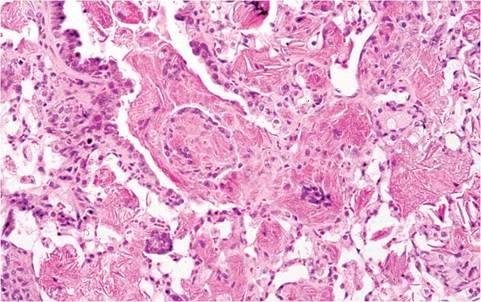
FIG. 1.104. Terminal airway of an aged B6 mouse with acidophilic
macrophage pneumonia. Hyaline, eosinophilic crystals are present within alveolar macrophages and alveolar spaces with multinucleate giant cell formation.
They are present in alveolar spaces, alveolar ducts, terminal airways, and bronchiolar glands. Crystal-bearing multinucleated giant cells and granulocytes are frequently scattered within affected areas of the lung. The crystalline material is complex and has been shown to contain iron, alpha-1 antitrypsin, immunoglobulin, and breakdown products of granulocytes. Based on ultra- structural studies, the crystals resemble Charcot-Leyden crystals, which are unique to humans and nonhuman primates in association with eosinophil-related diseases. AMP crystals are composed predominantly of Ym1 chitinase. Any disease process that impairs normal clearance (pulmonary tumors, Pneumocystosis, or other chronic pneumonias) can predispose to AMP.
AMP may be very extensive, leading to dyspnea in some mice.Although AMP is the most overt manifestation of this condition, "hyalinosis" of olfactory, nasal respiratory, middle ear, trachea, lung, stomach, gall bladder, bile duct, and pancreatic duct epithelium is part of the syndrome. Hyaline eosinophilic material fills the cytoplasm of affected epithelium, with blebbing of material from their apical surfaces (Fig. 1.105). Square to
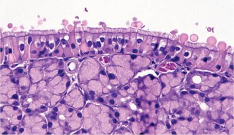
FIG. 1.105. Nasal epithelium from a B6 mouse, depicting hyaline eosinophilic material in the cytoplasm of respiratory epithelium and blebbing from the apical surfaces.
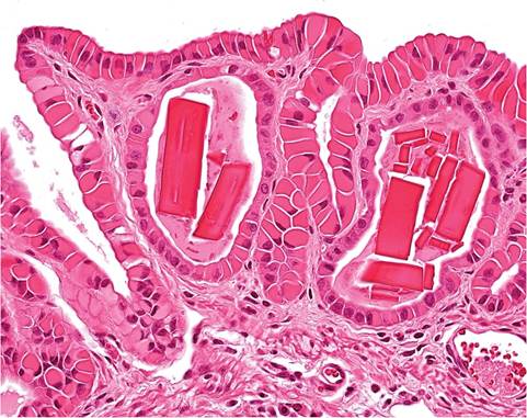
FIG. 1.106. Gall bladder of a B6 mouse with hyaline eosinophilic
material in biliary epithelial cells and crystals within mucosal glands.
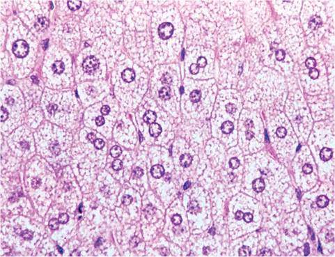
FIG. 1.107. Liver from a case of Reye's-like syndrome in a BALB/ cByJ mouse. Hepatocytes are distended with microvesicles and compress sinusoidal spaces.
rhomboid extracellular crystals may accumulate in glands of these tissues (Fig. 1.106). As in the lung, the hyaline material is composed of Ym1 and Ym2 chitinase. These changes may lead to thickening of the bile ducts and gall bladder, with dilatation and thickened, opaque walls.