Anatomy and physiology
Anatomy (derived from the Greek words meaning "to cut open") is the study of the morphology, or structure, of organisms. Thus, strictly speaking, anatomy deals with form rather than function.
It can be divided into macroscopic (gross) or microscopic anatomy. Macroscopic anatomy deals with structure that can be seen with the naked eye, whereas microscopic anatomy deals with structure that can only be seen with the aid of a microscope. It is also important to appreciate that it is also much more than simply looking at smears of cells or stained tissue sections. Use of immunocytochemistry, fluorescence-labeled markers, multispec- tral cameras and sophisticated imaging software, and so on is revolutionizing our understanding of cell and tissue biology. Figure 1.1 provides an example of the ability to localize specific proteins within various cells of the mammary gland.Macroscopic anatomy can be approached in different ways. Regional anatomy, as the name implies, deals with all the structures, such as nerves, bones, muscles, and blood vessels, in a defined region such
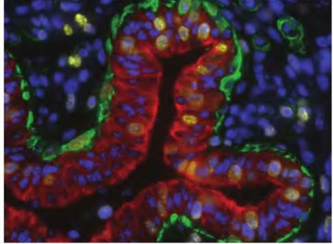
Fig. 1.1. Photomicrograph of a developing mammary duct. Taken from a Holstein calf, this tissue section was stained with specific antibodies and fluorescent tags to detect cell nuclei (blue), Cytokeratin 18 (red, a marker specific for epithelial cells), CD10 (green, a marker of myoepithelial cells), and Ki67 (yellow, a protein produced in nuclei of cells that are about to divide). The tissue section is from a study to evaluate the effects of the ovary on ontogeny of myoepithelial cells in the bovine mammary gland. Image is courtesy of Dr. Steve Ellis, Clemson University.
as the head or hip. Systemic anatomy entails the study of a given organ system such as the muscular or skeletal system.
It also involves the study of organ systems that are groups of organs that work together for a specific function, such as the digestive or urinary system. Surface anatomy considers markings that are visible from the outside. These may include knowledge of the muscles, such as sternocleidomastoid muscle, as a landmark to find another structure, such as the carotid artery.Microscopic anatomy includes cytology and histology. Cytology is the study of the structure of individual cells that constitute the smallest units of life, at least in the sense of animal physiology. Histology is the study of tissues. Tissues are a collection of specialized cells and their products that perform a specific function. Tissues combine to form organs such as the heart, liver, and brain, and will be explained in greater detail in Chapter 4.
Developmental anatomy is the study of the changes in structure that occur throughout life. Embryology is a subdivision of developmental anatomy that traces the developmental changes prior to birth. Many systems of the body are not completely developed at birth, hence the need to continue to follow their development after parturition. Specific to farm mammals, understanding and management of the postnatal development of the mammary gland and reproductive system are essential for the success of dairies, cow / calf operations, flocks of sheep and goats, piggeries, and so on.
Physiology is the study of the function of living systems. While various systems will be presented separately throughout this book, it must be recognized that all systems work together to maintain the normal functioning of an animal. Therefore, the cardiovascular system does not work in isolation from the respiratory or nervous system, but instead they work in unison to coordinate the distribution of oxygen and removal of carbon dioxide throughout the body. As in anatomy, there are levels of complexity.
Cellular physiology is the study of how cells work.
This includes the study of events at the chemical, molecular, and genetic levels. Organ physiology includes the study of specific organs, that is, cardiac or ovarian. Systems physiology includes the study of the functions of specific systems such as the cardiovascular, respiratory, or reproductive systems.As you study anatomy and physiology, it will become apparent that structure and function have evolved to complement each other. The complementarity of structure and function is an essential concept. At multiple levels, a return to this fundamental idea will hasten your grasp of what sometimes seems to be an overwhelming amount of information and detail. But ultimately, the point is for you to understand how an animal works and to understand limitations. This relationship between form and function is evident beginning at the cellular level. For example, the epithelial cells that line the internal surface of the small intestine have so-called tight junctions that act to restrict the movement of materials into the body from the gastrointestinal tract, whereas the epithelial linings (endothelial cells) of capillaries have modified junctions. The linings of capillaries must be sufficiently porous to allow solutes to move readily in either direction across the capillary wall to nourish the tissue and remove waste products.
As another example, there are structural differences between birds and mammals that allow flight. Birds contain pneumatic bones, that is, bones that are hollow, which are connected to the respiratory system. These bones include the skull, humerus, clavicle, keel, sacrum, and lumbar vertebrae. In addition, the lumbar and sacral vertebrae are fused as an adaptation for flight. This provides yet another example of complementary structure and function.