Anatomy of a muscle
A classic' shaped muscle (Fig. 4.2) has a thick fleshy central part called the lx,lh∣. and tapers at each end - the head. Here, the connective tissue muscle sheath is continuous with the dense fibrous connective (issue of the tendon that attaches the muscle to a bone.
A muscle is attached to a bone at two points: its starting point is called its origin; this moves least during contraction. The opposite end. where the muscle inserts on the bone, is called the insertion. However, a muscle can have more than one belly, all inserting al one point, in which case the muscle is said to have a number of heads, e.g. the biceps muscle has two heads. The length of the tendon attaching the muscle to a bone will vary, and in some cases tendons are far longer than the muscle itself, e.g. Ilexor and extensor tendons running over the digits.Not all muscles take the 'classic' shape described above. Sometimes they are present in flat sheets, in
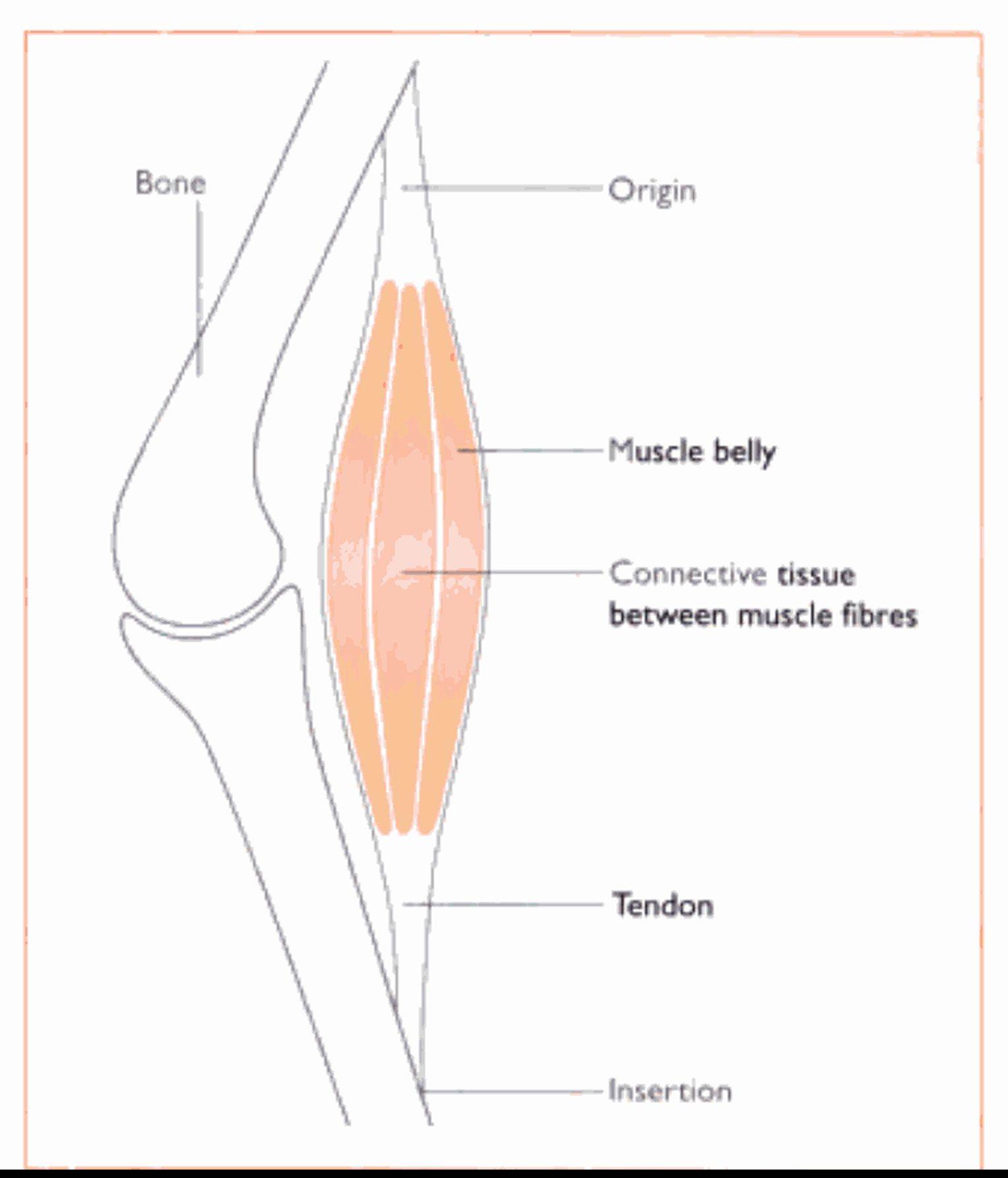
Fig. 4.2 Basic structure of a muscle The connective tissue between the muscle fibres is continuous with that of the tendon.
Muscles of mastication (Fig. 4.4)
The main muscles responsible for the masticatory or *chewing, action of the jaw arc:
Digastricus - this muscle opens the jaw. aided by gravity, and is Iocaled on lhe Caudovenlral surface of the mandible. Its origin is lhe jugular process of the occipital bone and it inserts on the angle and ventral surface of the mandible
Masseler - this muscle closes the jaw and lies lateral to the mandible. It originates from the zygomatic arch and inseris on the masseteric fossa on lhe lateral surface of lhe mandible Temporalis - this muscle also closes the jaw and is (he Iargesl and strongest muscle of lhe head. Il covers much of lhe dorsal and lateral surfaces of Ihe skull. Il Iills the temporal fossa of lhe skull and inserts on lhe coronoid process of the mandible Medial and lateral pterygoids - these are deep muscles that lie medial to Ihe mandible. They aid lhe temporalis and masseter muscles in closing lhe jaw. but they are also responsible for the side to side movements of lhe mouth.