Anatomy of the urinary system
The urinary system is critical for homeostasis and performs many essential functions. These include
• regulation of blood volume and blood pressure
• control of blood concentrations of several ions (e.g., Na, K, Ca)
• maintenance of blood pH via control of H+ and HCO3 ion secretion
• elimination of waste products and recovery of filtered nutrients.
The urinary system is composed of the paired kidneys and ureters, urinary bladder, and urethra. Essentially, dissolved materials in the liquid fraction of blood (plasma) are formed into a filtrate by the action of the kidneys. Once this filtrate is created, some additional materials are added (secretion), but others are recovered (reabsorption). The liquid that makes it through the microscopic tubular nephrons of the pelvis of the kidney and ultimately the urinary bladder exits the body as urine. Urine is typically slightly acidic (~pH6.0), but its volume and composition varies depending on metabolism, diet, and the need to produce either dilute or concentrated urine to maintain extracellular fluid volume and osmolarity.
Let's now consider some of the detailed anatomy of this system, beginning with the kidneys. WeTl use the ovine kidney to characterize some of the major features. In their normal retroperitoneal position, the kidneys are located between the 12th thoracic and third lumbar vertebra. They are held in place by the peritoneum and are in contact with adjacent visceral and surrounded by a layer of adipose tissue. Consequently, the kidneys are generally well protected. The outermost renal fascia anchors the kidney to the peritoneum, while the center layer of adipose tissue provides additional support and cushioning. The innermost connective tissue layer is the renal capsule. It is directly adjacent to the outer surface of the kidney parenchymal tissue.
The photograph provided in Figure 16.1 shows a bisected preserved sheep kidney. A portion of the thin but tough renal capsule is indicated. This specimen and companion drawing illustrate major macroscopic features of the kidney. However, as we'll soon see, the ultimate work of filtration and reabsorption is done by complex multicellular tubules called nephrons that are only evident at a microscopic level. These functional units of the kidney are located in lobules within distinct zones or regions of the kidney that can be grossly distinguished.Anatomy and Physiology of Domestic Animals, Second Edition. R. Michael Akers and D. Michael Denbow. © 2013 John Wiley & Sons, Inc. Published 2013 by John Wiley & Sons, Inc.
The outer region or zone, the cortex, lies over the inner region called the medulla and a central area called the renal pelvis or hilus. This central area is the location for the entrance and exit of the renal vein and artery as well as the origin of the ureter, which conducts urine to the bladder. The model illustrated in Figure 16.2 provides a three-dimensional represen-
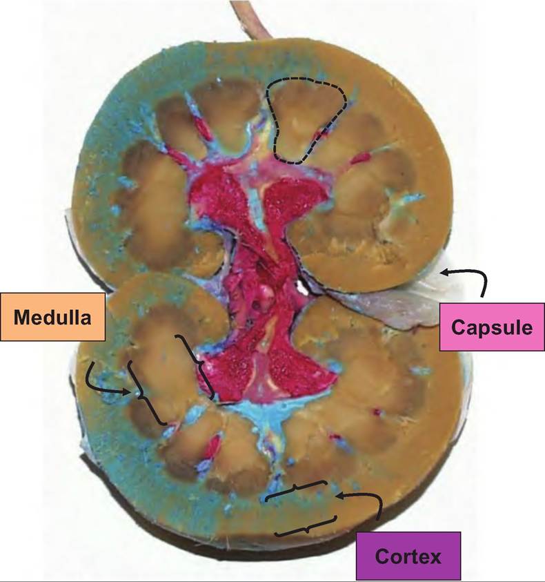
Fig. 16.1. Bisected preserved sheep kidney. Red latex fills much of the renal pelvis or hilus. The cortex is the outer rim of parenchymal tissue (brackets) and the medulla occurs in the region between the renal pelvis and cortex. A portion of the protective renal capsule is evident as a thin, membrane-like material. The dotted line outlines the region of a renal pyramid.
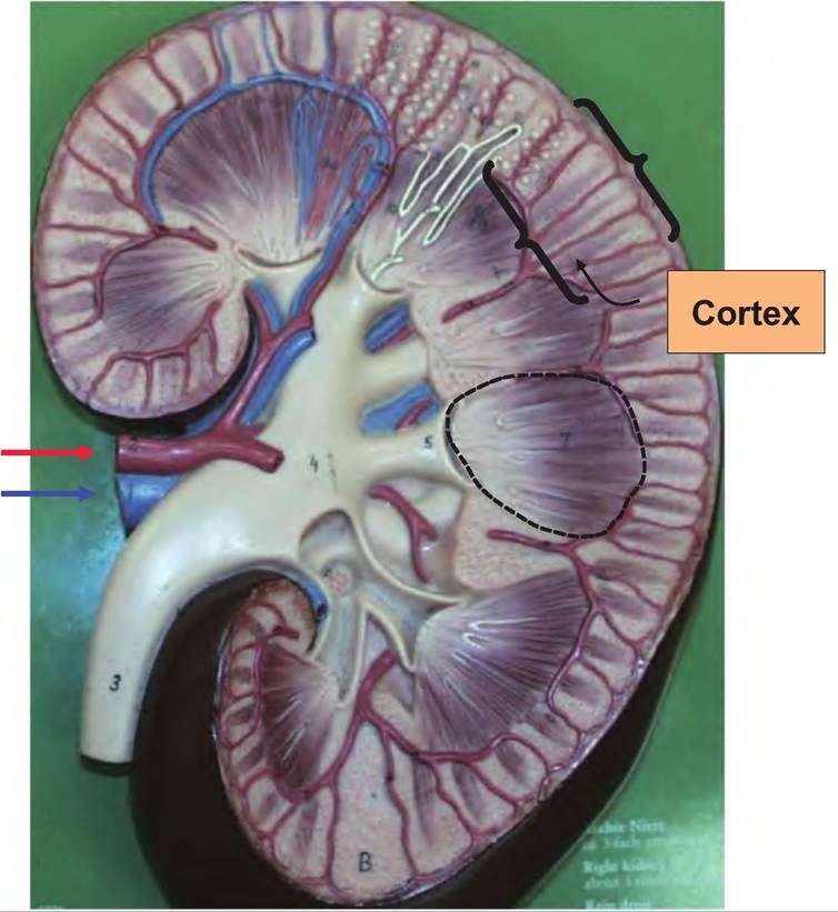
Fig. 16.2. Model of kidney. The model illustrated in this photograph demonstrates in greater detail gross anatomy of the kidney. The renal vein and artery (arrows) are evident in the region of the renal pelvis, as is the ureter (3). The funnel-like structures that feed the exiting ureter are the major (4) (closest to the ureter4) and minor calyces (5).
These structures capture filtrate from the tips of the renal pyramids as shown in the cutaway region. An example of a renal pyramid is illustrated by the dotted lines in Figure 1 6.1 and Figure 16.2.tation of these structures. Figure 16.3 gives a "flow" diagram to link blood flow and corresponding urine production in the kidney. Briefly, the renal artery and vein branch at nearly right angles to supply each of the kidneys. As each of these vessels approaches the hilus, they branch into smaller segmental arteries, named because they supply blood to sectors or segments of the mass of the kidney tissue. Each of the segmental arteries divides to create lobar arteries that divide to yield interlobar arteries that pass between the pyramids of the medulla toward the kidney cortex.
Near the boundary between the cortex and medulla the interlobar arteries branch into the arcuate arteries that arch (hence the name) over the bases of the medullary pyramids. Small interlobular arteries radiate outward to supply the tissue of the cortex. Most of the blood (~90%) that enters the kidney supplies the cortical tissue. Not surprisingly, this is the region where the bulk of the nephrons are located. The veins trace essentially the same pathway in reverse (Fig. 16.4).
Among the domestic species, swine and large ruminants have kidneys that are described as multipyrami- dal or multilobar. In these cases, a papilla (essentially
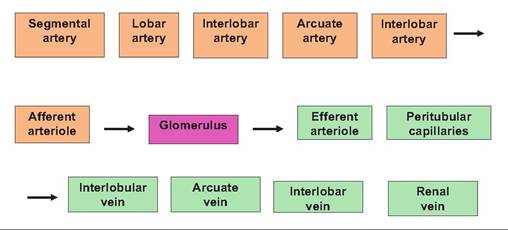
Fig. 16.3. Kidney blood flow. The group of progressive vessels (left to right) is the arterial branches (tan boxes) that ultimately supply the capillaries of the glomerulus where filtration takes place to supply fluid that enters the lumen of the nephron. Elements of the blood that are not filtered (cells and large proteins) and blood that is not subjected to filtration leaves the capillaries of the glomerulus and enters the venous circuit to exit the kidney via the renal vein (green boxes).
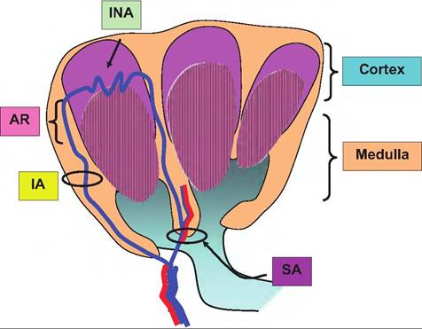
Fig. 16.4. Kidney blood supply. This stylized drawing illustrates some of the blood supply to kidney parenchymal tissue. SA, segmental artery; IA, interlobar arteries; AR, arcuate arteries; and INA, Interlobulararteries.
the tip of the pyramid) projects into the space of a minor calyx, and this is continuous with the ureters. Unipyramidal or unilobar kidneys occur in most carnivores, small ruminants, and horses. The kidney (of the cat, for example) consists of one lobe that results from the fusion of several lobes during development. A single broad ridge or crest created by the fusion of the papillae is intimately associated with an expanded internal portion of the ureters, which spreads over the internal surface of the renal pelvis (Banks, 1983).
Under usual circumstances, blood flow to the kidney is impressive, typically averaging 25% of cardiac output. As blood enters the renal artery, it progresses as outlined in Figure 16.3. The essential feature is that blood eventually passes into the tufts of capillaries that constitute the renal corpuscle (a surrounding structure called Bowman's capsule + the tuft of capillaries) that is connected with the first segment of the nephron (proximal convoluted tubule).