Nephron structure
The function of the kidney is tied to the nephrons. Each nephron consists of the glomerulus (the tuft of blood vessels) and glomerular capsule (often called Bowman's capsule). This tuft or knot of capillaries has an afferent (toward) and efferent (away) arteriole.
Blood that enters is subjected to filtration and osmotic pressures that act to force some of the liquid of the blood between the endothelial cells into the space surrounded by Bowman's capsule. Blood cells, larger molecules, and the remaining liquid exit the tuft of capillaries via the efferent arteriole. Liquid that passes out of the capillaries into the space of Bowman's capsule enters into the first segment of the tubular portion of the nephron called the proximal convoluted tubule (PCT). This segment of the nephron gets its name because the tube is very highly coiled (convoluted), and it is the closest to the site of filtrate formation (proximal).In sequence, the remaining parts of the nephron are the portion of the PCT leading to the thin or descending limb of the loop of Henle, ascending loop of Henle, distal convoluted tubule (DCT), and collecting duct (CD). The ends of the collecting ducts are located at the tips of the renal pyramids. This means that liquid that exits the nephrons at this point enters the ureter, passes to the bladder, and is lost as urine. Nephrons occur in two classes. The majority class (cortical) is located primarily within the kidney cortex. However, so-called juxtamedullary nephrons are arranged near the boundary between the cortex and medulla so that the loop of Henle for these nephrons passes deep into the medullary region. As we'll soon see, these nephrons play an especially important role in regulation of blood osmolarity. Figure 16.5 illustrates the orientation of nephrons within the kidney tissue.
In particular, notice that the branches from the arcuate arteries (paired artery and vein that arch over the boundary between the cortex and medulla) supply the interlobular arteries that ultimately supply the efferent arterioles of the glomerulus.
These appear as circular white balls in the photograph. The two classes of nephrons (cortical) and the longer juxtamedullary nephrons are also illustrated in this photograph. The large branched structure in the center illustrates a CD, which, as the name suggests, collects effluent from the DCT of numerous nephrons as it traverses along the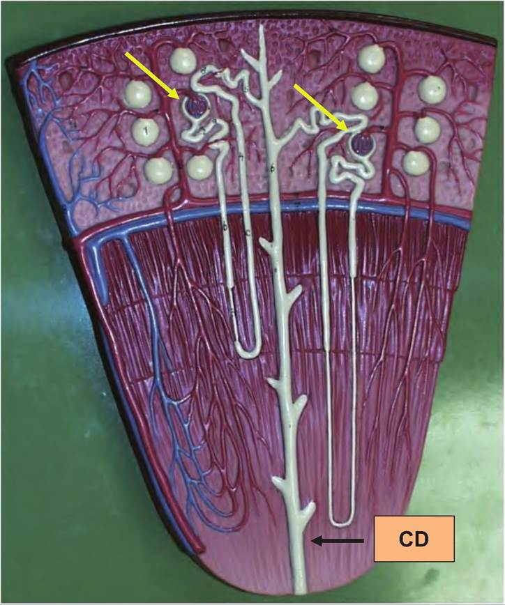
Fig. 16.5. Organization of nephrons. This photograph of a kidney model illustrates the orientation of microscopic structure within the tissue of a kidney pyramid. Numerous renal glomeruli (white globular structures) populate the renal cortex. Cross-sectioned glomeruli (arrows) show the funnel-like arrangement of the Bowman's capsule surrounding the tuft of capillaries and the afferent and efferent arterioles. The coiled tubule immediately exiting the glomerulus is the proximal convoluted tubule. Its path can be traced, as it becomes the descending then ascending loop of Henle. Once back in the region of the glomerulus, the tubular nephron becomes the distal convoluted tubule before it joins the collecting duct (CD).
length of the renal pyramid. Further detail of the structure of the glomerulus and initial segment of the nephron is shown in Figure 16.6.
Figure 16.7 provides a drawing to illustrate the components of a nephron and associated blood supply to the glomerulus. The mammalian kidney is the best- understood osmoregulatory organ in the animal kingdom, thanks to extensive research over the past 40 years. Activities linked with the mammalian kidney include a number of functions that are tied with other organs in lower vertebrates, for example, the skin, bladder, and gills of fishes or the salt glands of many reptiles and birds. This perhaps explains the serious nature of kidney disease or defects in our animals. In short, there is no substitute for a healthy, wellfunctioning urinary system. As we have indicated in prior sections, structure and function are closely allied.
The kidney and especially the elegant arrangement of the sections of the nephron, associated blood supply, and, finally, creation of a continuously maintained osmotic gradient within the tissue of the renal medulla are critical to kidney function. The nephron is a highly convoluted but nonetheless simple tube composed of a single layer of epithelial cells. The tube is essentially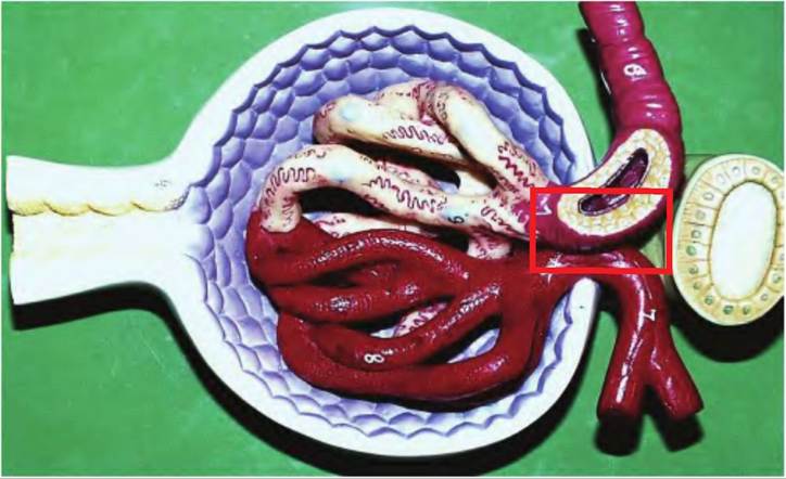
Fig. 16.6. Structure of the glomerulus. This photograph illustrates cellular structure associated with the glomerulus. The larger vessel on the upper right illustrates the afferent arteriole; the pale yellow covering on the surface of the vessel once it enters Bowman's capsule illustrates a layer of cells called podocytes that support the endothelial cells of the afferent capillary bed. These cells aid in regulation of the filtration process as discussed in the following. The other half of the tuft of vessels demonstrates the efferent vessels leading to the efferent arteriole. The pale blue layer of cells inside the Bowman's capsule represents the simple squamous epithelial cells that line its internal surface. To the left, these cells give rise to simple cuboidal epithelial cells that constitute the internal lining of the proximal convoluted tubule (CT). The structure on the extreme right illustrates a cross section of the distal CT. The boxed area represents cells of the wall of the afferent arteriole and adjoining distal CT that comprise a structure called the juxtaglomerular apparatus or JGA. As discussed in the text, the JGA is important in sensing decreases in blood pressure that lead to homeostatic events to restore pressure to normal.
closed because of the tuft of capillaries and filtered fluid as it leaves the Bowman's capsule, but it is open at its distal end as it joins the collecting ducts that ultimately empty into the renal pelvis. We will explore these relationships by discussing in some detail the cellular structure of different epithelial cells located along the course of the nephron. To understand the role of the kidney in long-term control of blood pressure, blood flow, and stimulation of erythrocyte production, we will describe the importance of a specialized cluster of modified distal convoluted cells called the macula densa and the juxtaglomerular cells of the wall of the afferent arteriole, which together make up the juxtaglomerular apparatus. In addition, we'll consider the significance of the network of peritubular capillaries that intertwine around the loop of Henle and the curved course of blood flow that mirrors the hairpin bend in the loop of Henle called the vasa recta.