BACTERIAL INFECTIONS
Bordetella bronchiseptica Infection
This bacterium is a potential problem for gerbils, but has not been reported as a natural disease. Young gerbils inoculated intranasally with B.
bronchiseptica developed severe disease with high mortality, while older gerbils appeared to be more resistant. Both M. unguiculatus and Meriones shawi were susceptible. Because of the frequency of B. bronchiseptica in laboratory guinea pigs and rabbits, contact of gerbils with these species should be avoided.Cilia-Associated Respiratory (CAR) Bacillus Infection
Gerbils are susceptible to experimentally induced infections with the CAR bacillus. Young gerbils inoculated intranasally with a rat isolate were subclinically infected during the study. However, at necropsy, there was colonization of the apices of epithelial cells lining the trachea and airways, with marked peritracheal and peribronchial lymphocytic infiltration. The relevance of these findings under natural conditions remains unclear.
Citrobacter rodentium Infection
An outbreak of diarrhea in a Spanish laboratory animal facility was attributed to infection with C. rodentium. The presenting signs were bloody diarrhea, rough hair coat, wasting, high mortality, and, on gross and microscopic examination, thickening of the colonic and rectal walls, with goblet cell metaplasia. Citrobacter rodentium was isolated from the large intestines of animals sampled during the acute stages of the disease.
Clostridium difficile Enterocolitis: Antibiotic Toxicity
Typhlocolitis with mortality has occurred in gerbils that received a combination of amoxicillin and metronidazole in their food. Deaths occurred beginning on day 7 following treatment, and typhlitis and colitis were the lesions observed at necropsy. Clostridium difficile was recovered on anaerobic culture, and C. difficile exotoxins were demonstrated by ELISA.
This is a complication following oral antibiotic treatment designed to eliminate naturally occurring Helicobacter spp. infections in gerbils.Clostridium piliforme Infection: Tyzzer's Disease
Mongolian gerbils are exquisitely susceptible to fatal Tyzzer's disease, caused by C. piliforme. There have been numerous documented cases of Tyzzer's disease in this species. Typically, the experimental induction of the disease in rodents requires treatment with immunosuppressive drugs, such as cortisone. However, Tyzzer's disease can be produced readily in gerbils without benefit of immunosuppression. Young gerbils have developed the typical disease following the oral inoculation of isolates from other species. Gerbils appear to be more susceptible to clinical disease following exposure to C. piliforme than do immunosuppressed mice. Gerbils caged with nonautoclaved soiled bedding suspected to be contaminated with the organism have been used as sentinels for detection of subclinical infections or environmental contamination with C. piliforme. Typical clinical signs associated with Tyzzer's disease in gerbils include depression, ruffled hair coat, hunched posture, anorexia, and watery diarrhea. Following oral inoculation, severely affected animals usually die within 5-7 days postinoculation. In addition to focal hepatic necrosis, bacterial antigen has been observed in ileocecal enterocytes by 3 days. Extensive lesions and bacterial antigen have been demonstrated in the jejunum, ileum, and cecum by 5-6 days. In affected gerbils, bacterial antigen may also be present in the muscle layers of the intestine and in Peyer's patches. Ileal enterocytes and Peyer's patches may be the initial sites for bacterial growth.
Pathology
Pinpoint, pale foci up to 2 mm in diameter are usually present in the liver. Ecchymoses on the small intestine and cecum are variable findings. The walls of the small intestine and cecum are usually edematous. Intestinal contents are fluid and sometimes contain blood.
The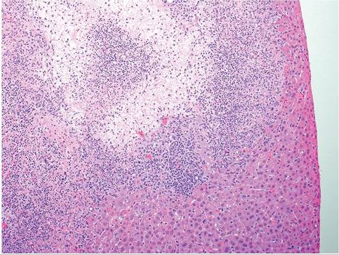
FIG. 4.2. Focal necrotizing hepatitis in a young Mongolian gerbil with acute Tyzzer's disease.
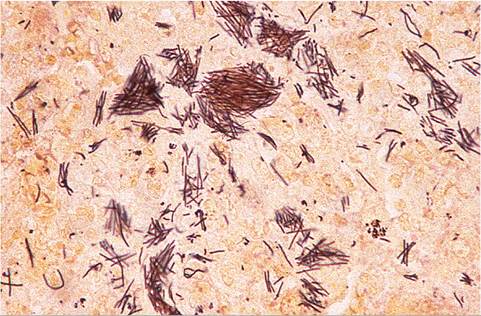
FIG. 4.4. Hepatic lesion from a Mongolian gerbil with Tyzzer's disease, stained with the Warthin-Starry method. Note the bundles of intracytoplasmic Clostridium piliforme bacilli in hepatocytes.
mesenteric lymph nodes may be enlarged and edematous. On microscopic examination, liver lesions are frequently concentrated in the periportal regions. In acute cases, there are foci of coagulation to caseation necrosis, with variable leukocytic infiltration, neutrophils predominating (Fig. 4.2). Intracytoplasmic bacilli are most numerous in hepatocytes adjacent to necrotic foci (Figs. 4.3 and 4.4). In hepatic lesions interpreted to be several days in duration, there may be focal fibrosis with mineralization. Intestinal lesions are usually most extensive in the ileum and cecum. Necrosis and sloughing of enterocytes, blunting of villi in affected areas, and transmural edema occur. Leukocytic infiltrates in the lamina propria consist of neutrophils and mononuclear cells. There may be necrosis of the adjacent intestinal smooth muscle, with leukocytic infiltration (Fig. 4.5).
Frequently focal necrosis of Peyer's patches and mesenteric lymph nodes occurs. Intracytoplasmic bacilli are usually evident in enterocytes and sometimes in smooth muscle cells. Myocardial lesions, when present, consist of focal coagulation necrosis, with collapse of myofibers, and leukocytic infiltration (Fig. 4.6). There may be mineralization of cell debris. Bundles of bacilli may be evident in cardiac myofibers bordering necrotic foci using Warthin-Starry or Giemsa stains. Diffuse suppurative encephalitis is another possible manifestation of Tyzzer's disease in this species.
Diagnosis
The presence of the typical gross and microscopic lesions and the histochemical demonstration of intracellular fascicles of bacilli are sufficient to confirm the diagnosis.
Differential diagnoses include bacterial enterocolitis associated with C. rodentium, C. difficile, and Salmonella spp. infections.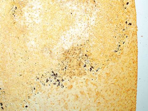
FIG. 4.3. Same region as the previous figure, stained with Warthin- Starry method. Note the argyrophilic Clostridium piliforme bacteria at the periphery of the necrosis.
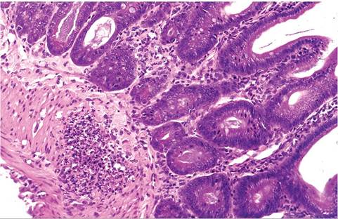
FIG. 4.5. Ileum from a Mongolian gerbil with Tyzzer's disease. Note the leukocytic infiltrate in the lamina propria and the focus of leukocytic infiltration in the adjacent muscularis.
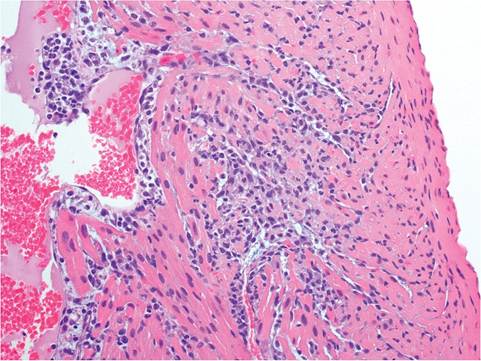
FIG. 4.6. Focal nonsuppurative myocarditis in a Mongolian gerbil with Tyzzer's disease.
Helicobacter spp. Infections
Naturally occurring enteric Helicobacter spp. infections have been identified in gerbils. In a survey conducted in Japan, Helicobacter hepaticus was detected in the feces of gerbils collected from several premises, Helicobacter bilis was detected in the feces of gerbils acquired from a commercial supplier in the United States, and Helicobacter rodentium has also been detected in inbred gerbils from Japan. Lesions and clinical signs were absent in these animals. Diagnosis can be achieved by culture or PCR of feces or intestine. Following experimental inoculation with Helicobacter pylori, Mongolian gerbils develop chronic gastritis, gastric ulcers, intestinal metaplasia, and gastric adenocarcinomas. The Mongolian gerbil is an established animal model for the study of gastric carcinogenesis associated with chronic H. pylori infection.
Leptospira spp. Infection: Leptospirosis
Leptospirosis has not been reported as a natural infection in gerbils, but gerbils are quite susceptible to experimental infection with a number of Leptospira serovars, although different stocks of gerbils may vary in susceptibility.
Acute disease is characterized by hemolytic icterus, with pale, mottled livers. Microscopically, there is degeneration of renal distal convoluted tubules and centrilobular hepatocytes with conspicuous erythropha- gocytosis in the spleen. Spirochetes are present in kidney and liver in large numbers. Chronic infection occurs frequently, with chronic nonsuppurative inflammation, interstitial fibrosis, and development of progressively severe tubular degeneration and cyst formation. The infection may persist in the kidney for months to years. Thus, leptospirosis remains a yet to be reported natural infection in gerbils, but has high potential as a disease to be discovered in gerbils.Listeria monocytogenes Infection: Listeriosis
Listeriosis occurs naturally in a number of rodents and lagomorphs. Natural infection of Mongolian gerbils with L. monocytogenes has not been reported, but a natural outbreak of listeriosis was observed in bushy-tailed jirds (Sekeetamys calurus). Affected animals died acutely with no prior clinical signs. Necrotizing lesions with large numbers of Gram-positive coccobacilli were present in liver, intestine, spleen, hepatic lymph nodes, and mesenteric lymph nodes. Most of the animals also had pneumonitis, with fewer numbers of visible bacteria. Diagnosis can be confirmed by culture, which is enhanced when tissues are preincubated at low temperatures prior to culture.
Salmonella enterica Infection: Salmonellosis
Disease and mortality have been observed in young gerbils 3-10 weeks of age that were naturally infected with S. enterica serovar Typhimurium. Clinical signs included moderate to severe diarrhea, dehydration, weight loss, and leukocytosis with neutrophilia. The mortality rate reached over 90%. In 1 report, animals also had a heavy infestation with Rodentolepis nana. An outbreak of salmonellosis in a gerbil colony due to a S. enterica serovar Enteritidis (O antigen Group D) has also been reported. Salmonella-infected cockroaches were implicated as a possible source of the infection.
The gastrointestinal tract is usually distended with gas and fluid ingesta. Fibrinopurulent exudate may be present in the peritoneal cavity in some animals. Microscopic hepatic lesions may vary from foci of leukocytic infiltration to larger foci consisting of central caseation necrosis with variable mineralization with epithelioid cells, lymphocytes, and neutrophils oriented around the periphery. Exudation of neutrophils into crypt lumina is occasionally present in the intestine. Focal hepatitis, splenic necrosis, suppurative orchitis, interstitial pneumonia, and purulent or pyogranulomatous leptomeningitis have been observed microscopically in gerbils. Salmonella may be cultured from sites such as the small intestine, liver, spleen, and heart blood. The primary differential diagnosis would be Tyzzer's disease.
Staphylococcus aureus Infection: Staphylococcal Dermatitis
Acute, diffuse dermatitis has been associated with beta- hemolytic S. aureus infection. The disease appears to affect primarily young gerbils, and there may be a relatively high morbidity and mortality. The disease was reproduced in gerbils inoculated in the nasal region with the staphylococcal isolate. On gross examination, there may be a diffuse moist dermatitis involving the face, nose, feet, legs, and ventral body surface. Alopecia, erythema, and moist brown exudate have been associated with the typical lesions. Microscopic changes are those of a suppurative dermatitis, with neutrophils infiltrating into the superficial and deep dermis and adnexae, with concurrent acanthosis and hyperkeratosis. Ulcerations may occur. Focal suppurative hepatitis may be present in some fatal cases of the disease. Staphylococcus aureus and Staphylococcus xylosus have been associated with the syndrome nasal dermatitis (sore nose), probably as opportunistic infections (see “Nasal Dermatitis”).
BIBLIOGRAPHY FOR BACTERIAL INFECTIONS
Bordetella bronchiseptica Infection
Winsser, J. (1960) A study of Bordetella bronchiseptica. Proceedings of the Animal Care Panel 10:87-104.
Cilia-Associated Respiratory Bacillus Infection
St. Claire, M.B., Besch-Williford, C.L., Riley, L.K., Hook, R.R., & Franklin, C.L. (1999) Experimentally-induced infection of gerbils with cilia-associated respiratory bacillus. Laboratory Animal Science 49:421-423.
Citrobacter rodentium Infection
de la Puente-Rodondo, V.A., Gutierrez-Martin, C.B., Perez-Martinez, C., del Blanco, N.G., Garcia-Iglesias, M.J., Perez-Garcia, C.C., & Rodriguez-Ferri, E.F. (1999) Epidemic infection caused by Citrobacter rodentium in a gerbil colony. Veterinary Record 145:400-403.
Clostridium difficile Infection
Bergin, I.L., Taylor, N.S., Nambiar, P.R., & Fox, J.G. (2005) Eradication of enteric Helicobacters in Mongolian gerbils is complicated by the occurrence of Clostridium difficile enterotoxemia. Comparative Medicine 55:265-268.
Clostridium piliforme Infection
Carter, G.R., Whitenack, D.L., & Julius, L.A. (1969) Natural Tyzzer's disease in Mongolian gerbils (Meriones unguiculatus). Laboratory Animal Care 19:648-651.
Gibson, S.V., Waggie, K.S., Wagner, J.E., & Ganaway, J.R. (1987) Diagnosis of subclinical Bacillus piliformis infection in a barrier- maintained mouse production colony. Laboratory Animal Science 37:786-791.
Motzel, S.L. & Gibson, S.V. (1990) Tyzzer's disease in hamsters and gerbils from a pet store supplier. Journal of the American Veterinary Medical Association 197:1176-1178.
Port, C.D., Richter, W.R., & Moize, S.M. (1971) An ultrastructural study of Tyzzer's disease in the Mongolian gerbil (Meriones unguiculatus). Laboratory Investigation 25:81-87.
Veazey, R.S., 2nd., Paulsen, D.B., & Schaeffer, D.O. (1992) Encephalitis in gerbils due to naturally occurring infection with Bacillus piliformis (Tyzzer's disease). Laboratory Animal Science 42:516-518.
Waggie, K.S., Ganaway, J.R., Wagner, J.E., & Spencer, T.H. (1984) Experimentally induced Tyzzer's disease in Mongolian gerbils (Meriones unguiculatus). Laboratory Animal Science 34:53-57.
Yokomori, K., Okada, N., Murai, Y., Goto, N., & Fujiwara, K. (1989) Enterohepatitis in Mongolian gerbils (Meriones unguiculatus) inoculated perorally with Tyzzer's organism (Bacillus piliformis). Laboratory Animal Science 39:16-20.
Helicobacter spp. Infections
Bergin, I.L., Taylor, N.S., & Fox, J.G. (1999) Helicobacter pylori- induced gastritis in U.S.-bred Mongolian gerbils. Contemporary Topics in Laboratory Animal Science 38:27.
Bergin, I.L., Taylor, N.S., Nambiar, P.R., & Fox, J.G. (2005) Eradication of enteric Helicobacters in Mongolian gerbils is complicated by the occurrence of Clostridium difficile enterotoxemia. Comparative Medicine 55:265-268.
Fox, J.G. & Wang, T.C. (2014) Dietary factors modulate Helicobacter- associated gastric cancer in rodent models. Toxicologic Pathology 42:162-181.
Goto, K., Ohashi, H., Takakura, A., & Itoh, T. (2000) Current status of Helicobacter contamination of laboratory mice, rats, gerbils, and house musk shrews in Japan. Current Microbiology 41:161-166.
Kodama, M., Murakami, K., Sato, R., Okimoto, T., Nishizono, A., & Fujioka, T. (2005) Helicobacter pylori-infected animal models are extremely suitable for the investigation of gastric carcinogenesis. World Journal of Gastroenterology 45:7063-7071.
Watanabe, T., Tada, M., Nagai, H., Sasaki, S., & Nakao, M. (1998) Helicobacter pylori infection induces gastric cancer in Mongolian gerbils. Gastroenterology 115:642-648.
Whary, M.T. & Fox, J.G. (2004) Natural and experimental Helicobacter infections. Comparative Medicine 54:128-158.
Leptospira spp. Infection
Lewis, C. & Grey, J.E. (1961) Experimental Leptospirapomona infection in the Mongolian gerbil (Meriones unguiculatus). Journal of Infectious Diseases 109:194-204.
Tripathy, D.N. & Hanson, L.E. (1976) Some observations on chronic leptospiral carrier state in gerbils experimentally infected with Leptospira grippotyphosa. Journal of Wildlife Diseases 12:55-58.
Yamada, M. (1991) Differential susceptibility of two stocks of Mongolian gerbils (Meriones unguiculatus) to Leptospira. Journal of Experimental Animal Science 34:1-5.
Listeria monocytogenes Infection
Tappe, J.P., Chandler, F.W., Westrom, W.K., Liu, S.K., & Dolensek, E.P. (1984) Listeriosis in seven bushy-tailed jirds. Journal of the American Veterinary Medical Association 185:1367-1370.
Salmonella enterica Infection
Clark, J.D., Shotts, E.B., Jr., Hill, J.E., & McCall, J.W. (1992) Salmonellosis in gerbils induced by a nonrelated experimental procedure. Laboratory Animal Science 42:161-163.
Olson, G.A., Shields, R.P., & Gaskin, J.M. (1977) Salmonellosis in a gerbil colony. Journal of the American Veterinary Medical Association 171:970-972.
Staphylococcus aureus Infection
Peckham, J.C., Cole, J.R., Chapman, W.A., Jr., Malone, J.B. Jr., McCall, J.W., & Thompson, P.E. (1974) Staphylococcal dermatitis in Mongolian gerbils (Meriones unguiculatus). Laboratory Animal Science 24:43-47.