PARASITIC DISEASES
Gerbils are experimentally susceptible to a wide variety of protozoal, helminth, and arthropod parasites that are not naturally indigenous to gerbils. Those with potential for natural infection of gerbils, through exposure to other laboratory rodents or through management practices, are covered in this section.
Protozoal Infections
Although natural cryptosporidiosis has not been observed in gerbils, infant and adult gerbils are experimentally susceptible to infection with a number of Cryptosporidium species without immunosuppression, including Cryptosporidium parvum, Cryptosporidium muris, and Cryptosporidium andersoni. Cryptosporidium parvum infects the small intestine and biliary epithelium, and C. muris and C. andersoni infect gastric mucosa. Lesions include mild mucosal hyperplasia with attachment of organisms to epithelium. Giardiasis has not been reported as a natural disease in gerbils, but gerbils are highly susceptible to infection with Giardia cysts of human origin (aka Giardia lamblia, Giardia intestinalis, and Giardia duodenalis). Trophozoites can be found in the upper small intestine, and heavy infections may occur throughout the bowel. Mild mucosal hyperplasia and increased mucin production were evident in infected gerbils. Nonpathogenic enteric protozoa that have been observed in laboratory gerbils include Tritrichomonas caviae and Entamoeba sp.
Helminth Infestations
Gerbils can become infested with several oxyurid nematodes, but none appear to cause clinical problems. Dentostomella translucida has been reported in a variety of gerbils (Fig. 4.7). It may inhabit both the small and large intestine, and is notably larger than other rodent pinworms. Gerbils are also susceptible to contact infestation with the mouse and rat pinworms Syphacia obve- lata, Aspiculuris tetraptera, and Syphacia muris. Severe infestations with the “dwarf tapeworm” (R.
nana) have been reported in pet gerbils. Debilitation, dehydration, and mucoid diarrhea were presenting signs. In another report describing an epizootic of salmonellosis in Mongolian gerbils, affected animals were also heavily parasitized with R. nana. At necropsy, small tapeworms are present in the small intestine. On microscopic examination of smears of intestinal mucosa, or of paraffin- embedded sections of small intestine, eggs and cysticer- coids are readily identified. In view of the direct life cycle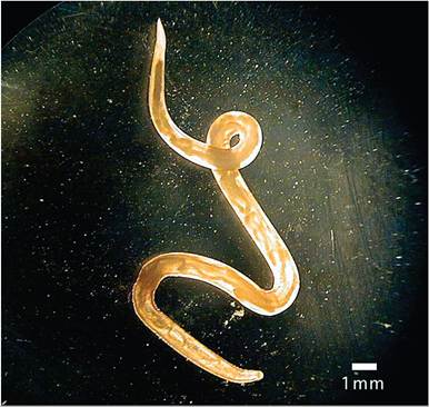
FIG. 4.7. Dentostomella translucida pinworm from the small intestine of a Mongolian gerbil. (Source: Wilkerson et al. 2010. Reproduced with permission from American Association for Laboratory Animal Science.)
of R. nana, there is a risk of transmission to human contacts. Hymenolepis diminuta has also been identified at necropsy in gerbils.
Arthropod Infestations
Mite Infestations: Acariasis
Gerbils can be hosts for infestation with Demodex spp., but clinical demodicosis is not considered to be a problem in healthy gerbils. The name Demodex meriones has been proposed, but it may represent Demodex aurati or Demodex criceti (hamster mites), since mites resembling both of these species have been found on the gerbil. Demodex mites were demonstrated in skin scrapings from a 4-year-old gerbil with diarrhea, cachexia, and rough hair coat. A lesion on the tail head was characterized by scaliness, hyperemia, and focal ulcerations. Old age and debilitation were considered to be important predisposing factors. There have been documented cases of skin lesions attributed to infestation with free-living nymphal astigmatic mites, Acarus farris infestations in gerbils. Copra itch mites (Tyrophagus castellani), probably introduced through the food, have been found incidentally on gerbils. Liponyssoides sanguineus, an ectoparasite occasionally seen in house mice, has also been observed in Mongolian and Egyptian gerbils.
Mites were also identified on laboratory mice and wild house mice on the same premises. No manifestations of disease were observed in affected animals. Mites were also present in the bedding in the cages.BIBLIOGRAPHY FOR PARASITIC DISEASES
Araujo, N.S., Mundim, M.J., Gomes, M.A., Amorim, R.M., Viana, J.C., Queiroz, R.P., Rossi, M.A., & Cury, M.C. (2008) Giardia duodenalis: pathological alterations in gerbils, Meriones unguiculatus, infected with different dosages of trophozoites. Experimental Parasitology 118:449-457.
Belosevic, M. (1983) Giardia lamblia infections in Mongolian gerbils: an animal model. Journal of Infectious Diseases 147:222-226.
Jacklin, M.R. (1997) Dermatosis associated with Acarus farris in gerbils. Journal of Small Animal Practice 38:410-411.
Kellogg, H.S. & Wagner, J.E. (1982) Experimental transmission of Syphacia obvelata among mice, rats, hamsters and gerbils. Laboratory Animal Science 32:500-501.
Kvac, M., Sak, B., Kvetonova, D., & Secor W.E. (2009) Infectivity of gastric and intestinal Cryptosporidium species in immunocompetent Mongolian gerbils (Meriones unguiculatus). Veterinary Parasitology 163:33-38.
Levine, J.F. & Lage, A.L. (1984) House mouse mites infesting laboratory rodents. Laboratory Animal Science 34:393-394.
Lussier, G. & Loew, F.M. (1970) Natural Hymenolepis nana infection in Mongolian gerbils (Meriones unguiculatus). Canadian Veterinary Journal 11:105-107.
Pinto, R.M., Gomes, D.C., & Noronha, D. (2003) Evaluation of coinfection with pinworms (Aspiculuris tetraptera, Dentostomella translucida, and Syphacia obvelata) in gerbils and mice. Contemporary Topics in Laboratory Animal Science 42:46-48.
Ross, C.R., Wagner, J.E., Wightman, S.R., & Dill, S.E. (1980) Experimental transmission of Syphacia muris among rats, mice, hamsters and gerbils. Laboratory Animal Science 30:35-37.
Schwartzbrott, S.S., Wagner, J.E., & Frisk, C.S. (1974) Demodicidosis in the Mongolian gerbil (Meriones unguiculatus): a case report. Laboratory Animal Science 24:666-668.
Vincent, A.L., Porter, D.D., & Ash, L.R. (1975) Spontaneous lesions and parasites of the Mongolian gerbil, Meriones unguiculatus. Laboratory Animal Science 25:711-722.
Wightman, S.R., Pilitt, P.A., & Wagner, J.E. (1978) Dentostomella translucida in the Mongolian gerbil (Meriones unguiculatus). Laboratory Animal Science 28:290-296.
Wightman, S.R., Wagner, J.E., & Corwin, R.M. (1978) Syphacia obvelata in the Mongolian gerbil (Meriones unguiculatus): natural occurrence and experimental transmission. Laboratory Animal Science 28:51-54.
Wilkerson, J.D., Brooks, D.L., Derby, M., & Griffey, S.M. (2001) Comparison of practical treatment methods to eradicate pinworm (Dentostomella translucida) infections from Mongolian gerbils (Meroines unguiculatus). Contemporary Topics in Laboratory Animal Science 40:31-36.
More on the topic PARASITIC DISEASES:
- Bibliography for parasitic diseases
- BIBLIOGRAPHY FOR PARASITIC DISEASES
- BIBLIOGRAPHY FOR PARASITIC DISEASES
- BIBLIOGRAPHY FOR PARASITIC DISEASES
- Content
- Clostridial Diseases
- GENERAL INTRODUCTION
- neurocysticercosis
- other Poxviral diseases
- THE CLOSTRIDIAL DISEASES