GENETIC, METABOLIC, AND OTHER DISORDERS
Epilepsy
Epileptiform seizures are common among Mongolian gerbils that are subjected to stress, which may include cage changing. Susceptibility begins at around 2 months of age and can reach an incidence of 40-80% within 6-10 months and persist throughout life.
The trait is inherited as a single autosomal locus with at least 1 dominant allele, with variable penetrance. The incidence, therefore, varies with different populations or lines of gerbils. Seizure-sensitive and seizure-resistant strains have been selected for experimental purposes. Clinical signs include twitching of vibrissae and pinnae, motor arrest, myoclonic jerks, clonic-tonic seizures, vestibular aberrations, and occasionally death. The dentate gyrus is believed to be the epileptic focus. Histopathologic lesions are not obvious.Nasal Dermatitis
“Sore nose,” also known as “red nose,” is a frequent problem in juvenile and adult Mongolian gerbils, appearing to be the most common in postpuberal animals. Nasal dermatitis is characterized by dermatitis and alopecia around the external nares and upper labial region. The incidence of the disease in individual colonies may be over 15%, but an incidence of around 5% is more typical. Mechanical trauma may contribute to the disease in some circumstances, but porphyrin-containing lacrimal gland secretions have been shown to be an important contributing factor. Secretions from the Har- derian gland normally bathe the eye and conjunctival sac and then are transported down the nasolacrimal duct to the external nares. The secretions are mixed with saliva and spread widely over the pelage during grooming. However, if these secretions are not removed routinely from the collection site at the external nares, chemical irritation and subsequent dermatitis may occur. Marked improvement has been seen in gerbils housed on sand, allowing animals to “sand bathe.” The
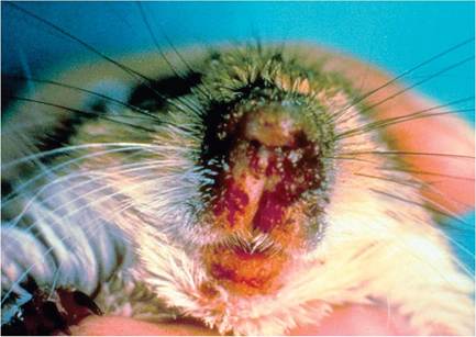
FIG.
4.8. Nasal dermatitis in mature Mongolian gerbil. There is marked reddening with serosanguinous encrustations around the external nares. (Source: © M.E. Olson.)failure to groom properly results in the accumulation of protoporphyrin-containing secretions around the external nares, resulting in local irritation, scratching, hair loss, and dermatitis. Intact gerbils fitted with Elizabethan collars, which prevented self-grooming, developed nasal dermatitis, while those with bilateral Harderian gland adenectomy did not develop the disease. Secondary infection with opportunistic bacteria such as S. xylosus or S. aureus may contribute to development of the moist, ulcerative form of the disease.
There may be varying degrees of dermatitis and alopecia involving the lateral and superior nasal area and the upper and lower lips (Fig. 4.8). Lesions may progress to severe ulcerative dermatitis, with exudation and excoriation and crusting in the upper labial region. Dermatitis and hair loss may also be present on the forepaws and periocular regions. Microscopically, there is hyperkeratosis and epidermal hyperplasia, with increased melanin deposition in the dermis. Epidermal spongiosis, hyperplasia, and necrosis, with infiltration and exudation of neutrophils, may be present in acute lesions. Other changes may include ulceration and epidermal abscessation.
Barbering
Conspecific hair chewing and plucking tends to be manifest as focal alopecia over the dorsum of the tail.
Tail Slip
Inappropriate restraint of gerbils may result in degloving of the tail skin. Gerbils should never be caught and held by the tip of the tail, but rather handled at the base of the tail only.
Periodontal Disease and Dental Caries
Gerbils that are maintained on a standard laboratory pelleted diet and water may develop progressively severe periodontal disease, which is first manifested at around 6 months of age and is readily apparent by 1 year.
Advanced disease is present in gerbils over 2 years of age, often with tooth loss.
They are also prone to the development of dental caries, which can be enhanced with cariogenic diets.Malocclusion
Lack of opposing occlusal contact results in elodont tooth overgrowth in all species of rodents, including gerbils. Reported cases in gerbils are rare and have been due to loss of the upper incisors with overgrowth of the lower teeth. Molar teeth of gerbils do not grow continuously.
Ocular Proptosis
Aged gerbils may develop protrusion of the nictitating membrane and conjunctiva with bulbar proptosis. The underlying cause has not been characterized.
Aural Cholesteatoma
Aural cholesteatomas, despite their name, are not neoplasms, but are rather concentric accumulations of keratinized epithelium. Spontaneous aural cholesteatomas occur in high frequency among adult gerbils, with a prevalence of over 50% at 2 years of age. These masses of keratinized epithelium arise from the outer surface of the tympanic membrane and external auditory canal (Fig. 4.9). As keratin is accumulated, it displaces the tympanic membrane into the middle ear. Compression and secondary inflammation result in the destruction of temporal bone and inner ear structures. Clinical signs include head tilt and accumulation of keratin plugs in the external ear canal. Differential diagnosis includes otitis media/interna, but this is rare in gerbils because of the vertical configuration of their Eustachian tubes.
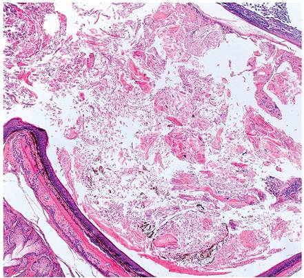
FIG. 4.9. Cholesteatoma in the external ear canal of a Mongolian gerbil. Note the keratin plug filling the canal.
Spongiosis of the Caudate Nucleus
Spongiform lesions arise in the auditory system of Mongolian gerbils, increasing in size, number, and extent with age. Lesions are most severe in the posterior ventral cochlear nucleus and the ventrolateral region of the caudal anterior ventral cochlear nucleus. The spongiosis is associated with neuronal necrosis and degeneration of axons, dendrites, and glia, but neuronal loss is subtle.
These lesions arise in laboratory gerbils, but are not apparent in progeny of wild-trapped gerbils. The clinical significance of these lesions is minimal.Focal Myocardial Degeneration
Focal myocardial degeneration and fibrosis are common microscopic findings in older gerbils. In general, 50% or more of male breeders may be affected, and a smaller percentage of breeding females. Lesions are probably ischemic in origin, but the etiopathogenesis has not been elucidated. On microscopic examination, there are foci of degeneration of myofibers, with interstitial fibrosis (Fig. 4.10).
Obesity and Diabetes
Approximately 10% of gerbils maintained on a standard laboratory diet can become obese. This condition can be associated with reduced glucose tolerance, elevated insulin, and hyperplastic or degenerative changes in the endocrine pancreas.
Hyperadrenocortism/Cardiovascular Disease of Breeding Gerbils
A disease complex, attributed to hyperadrenocorticism, has been described in repeatedly bred male and female, but not virgin, gerbils. Breeding females, and to a lesser extent males, develop mild to severe plaques of intimal and medial ground substance alterations with mineralization in the aorta and mesenteric, renal, and peripheral arteries. Breeders may have grossly visible plaques of the abdominal aorta, as well as aortic arch and the entire aorta in severe cases. Breeding animals have elevated serum triglycerides, enlarged pancreatic islets, fatty
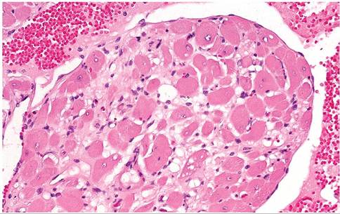
FIG. 4.10. Cardiac papillary muscle from an aged Mongolian gerbil with myocardial degeneration and interstitial fibrosis.
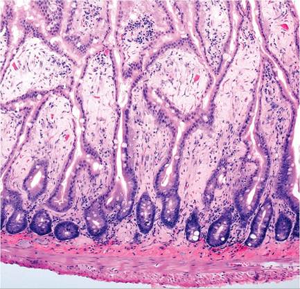
FIG. 4.11. Amyloid in the lamina propria of the small intestine in a Mongolian gerbil.
livers, thymic involution, adrenal hemorrhage, and adrenal lipid depletion. Some may have pheochromocytomas.
Male breeders have been found to have a high incidence of focal myocardial necrosis and fibrosis. This phenomenon is also linked to diabetes and obesity. Cause and effect relationships have not been firmly established, but it is clear that these lesions occur frequently in gerbil populations and seem to occur in higher prevalence among breeders. Spontaneously occurring stroke may be observed in aged animals.Amyloidosis
Amyloidosis can occur in aging gerbils, and has been reported in gerbils experimentally infected with a filariid worm. Clinical signs, when present, include weight loss, dehydration, anorexia, and death. Small intestinal lamina propria, liver, spleen, and lymph nodes (Figs. 4.11 and 4.12) are common sites of amyloid deposition. In
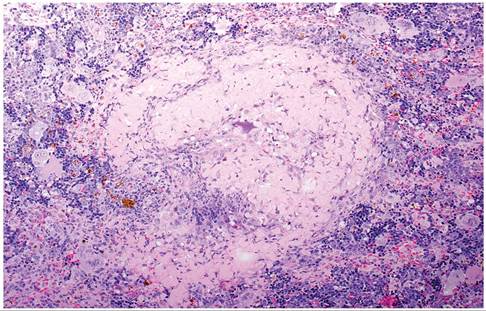
FIG 4.12. Amyloid in a splenic follicle of a Mongolian gerbil. Also note the prominent extramedullary hematopoiesis in the surrounding red pulp.
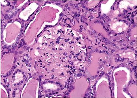
FIG. 4.13. Chronic progressive glomerulonephropathy in an aged Mongolian gerbil. There is thickening of glomerular and tubular basement membranes, with proteinaceous casts within tubules.
one study, the majority of cases were secondary to chronic renal disease.
Chronic Glomerulonephropathy
Glomerular hypercellularity, thickening of glomerular basement membranes, and tubular degeneration with dilation and cast formation in tubules are changes seen in the kidneys of aging gerbils (Fig. 4.13). Mononuclear cell infiltration consistent with chronic interstitial nephritis may be present in affected kidneys.
Cystic Ovaries
Female gerbils are prone to the development of ovarian cysts. Nearly 50% of gerbils over 400 days of age may be affected. Cysts range in size from 1 to 50 mm in diameter. Microscopic descriptions suggest that they are of follicular origin.
Ovulation and corpus luteum formation continue to occur in the presence of cysts, but litter sizes are reduced, and severely affected females become infertile.Toxic Disorders
Streptomycin Toxicity
Aminoglycoside antibiotics (dihydrostreptomycin, neomycin) cause a direct neuromuscular blocking effect at excessive doses by inhibition of acetylcholine release. Although other rodents and rabbits are susceptible to this effect, they are less likely to be treated with these drugs and are, in addition, big enough to receive the proper dose. The margin of safety for streptomycin is low, and antibiotic preparations are seldom prepared so that an appropriate dose in a small volume can be administered to a rodent. Gerbils treated with these preparations have developed acute toxicity, characterized by depression, ascending flaccid paralysis, coma, and death within minutes of administration.
Lead Toxicity
Because of their urine-concentrating ability, gerbils are prone to accumulation of lead and chronic lead toxicity. Their ability to accumulate lead in their kidneys is 4-6 times greater than rats. They are used for this purpose experimentally, and there is the potential for natural toxicity because of their gnawing behavior. Chronically toxic animals become emaciated. Their livers become small and pigmented; their kidneys pale and pitted. Microscopic findings include acid-fast intranuclear inclusions in proximal convoluted tubular epithelium and chronic progressive nephropathy. Occasional intranuclear inclusions may be found in liver, but the predominant finding is lipofuscin pigment granules in hepatocytes and Kupffer's cells. Gerbils may also develop a microcytic, hypochromic anemia with basophilic stippling. Differential diagnoses should include age-related glomerulonephropathy and erythrocytic basophilic stippling, a condition that occurs normally in the gerbil, but to a lesser degree.
Bibliography for genetic, metabolic,
AND OTHER DISORDERS
Bingel, S.A. (1995) Pathologic findings in an aging Mongolian gerbil (Meriones unguiculatus) colony. Laboratory Animal Science 45:597-600.
Marston, J.H. & Chang, M.C. (1965) The breeding, management and reproductive physiology of the Mongolian gerbil, Meriones unguiculatus. Laboratory Animal Care 15:34-48.
Norris, M.L. & Adams, C.E. (1972) Incidence of cystic ovaries and reproductive performance in the Mongolian gerbil, Meriones unguiculatus. Laboratory Animals 6:337-342.
Vincent, A.L., Porter, D.D., & Ash, L.R. (1975) Spontaneous lesions and parasites of the Mongolian gerbil, Meriones unguiculatus. Laboratory Animal Science 25:711-722.
Vincent, A.L., Rodrick, G.E., & Sodeman, W.A., Jr. (1979) The pathology of the Mongolian gerbil (Meriones unguiculatus): a review. Laboratory Animal Science 29:645-651.
Epilepsy
Buckmaster, P.S. & Wong, E.H. (2002) Evoked responses of the dentate gyrus during seizures in developing gerbils with inherited epilepsy. Journal of Neurophysiology 88:783-793.
Loskota, W.J., Lomax, P., & Rich, S.T. (1974) The gerbil as a model for the study of the epilepsies: seizure patterns and ontogenesis. Epilepsia 15:109-119.
Theissen, D.D., Lindzey, G., & Friend, H.C. (1968) Spontaneous seizures in the Mongolian gerbil (Meriones unguiculatus). Psycho- nomic Science 11:227-228.
Nasal Dermatitis
Breshnahan, J.F., Smith, G.D., Lentsch, R.H., Barnes, W.G., & Wagner, J.E. (1983) Nasal dermatitis in the Mongolian gerbil. Laboratory Animal Science 33:258-263.
Donnelly, T.M. (1997) Nasal lesions in gerbils (what's your diagnosis?). Laboratory Animals 27(2):17-18.
Farrar, P.L., Opsomer, M.J., Kocen, J.A., & Wagner, J.E. (1988) Experimental nasal dermatitis in the Mongolian gerbil: effect of bilateral Harderian gland adenectomy on development of facial lesions. Laboratory Animal Science 38:72-76.
Solomon, H.F., Dixon, F.M., & Pouch, W. (1990) A survey of staphylococci isolated from the laboratory gerbil. Laboratory Animal Science 40:316-318.
Theissen, D.D. & Kittrell, E.M.W. (1980) The Harderian gland and thermoregulation in the gerbil (Meriones unguiculatus). Physiology and Behavior 24:417-424.
Theissen, D.D. & Pendergrass, M. (1982) Harderian gland involvement in facial lesions in the Mongolian gerbil. Journal of the American Veterinary Medical Association 181:1375-1377.
Dental/Periodontal Disease
Afonsky, D. (1957) Dental caries in the Mongolian gerbil. New York State Dental Journal 23:315-316.
Fitzgerald, D.B. & Fitzgerald, R.J. (1965) Induction of dental caries in gerbils. Archives of Oral Biology 11:139-140.
Loew, F.M. (1967) A case of overgrown mandibular incisors in a Mongolian gerbil. Laboratory Animal Care 17:137-139.
Moskow, B.S., Wasserman, B.H., & Rennert, M.C. (1968) Spontaneous periodontal disease in the Mongolian gerbil. Journal of Periodontal Research 3:69-83.
Aural Cholesteatoma
Chole, R.A., Henry, K.R., McGinn, M.D. (1981) Cholesteatoma: spontaneous occurrence in the Mongolian gerbil, Meriones unguiculatus. American Journal of Otolaryngology 2:204-210.
Henry, K.R., Chole, R.A., & McGinn, M.D. (1983) Age-related increase of spontaneous aural cholesteatoma in the Mongolian gerbil. Archives of Otolaryngology 109:19-21.
Spongiosis of the Caudate Nucleus
McGinn, M.D. & Faddis, B.T. (1998) Neuronal degeneration in the gerbil brainstem is associated with spongiform lesions. Microscopy Research and Techniques 41:187-204.
Ostapoff, E.M. & Morest, D.K. (1989) A degenerative disorder of the central auditory system of the gerbil. Hearing Research 37:141-162.
Statler, K.D., Chamberlin, S.C., Slepecky, N.B., & Smith, R.L. (1990) Development of mature microcystic lesions in the cochlear nuclei of the Mongolian gerbil, Meriones unguiculatus. Hearing Research 50:275-288.
Metabolic Disease
Boquist, L. (1972) Obesity and pancreatic islet hyperplasia in the Mongolian gerbil. Diabetologia 8:274-282.
Nakama, K. (1977) Studies on diabetic syndrome and influences of long-term tolbutamide administration in Mongolian gerbils (Meriones unguiculatus). Endocrinologia japonica 24:421-433.
Wexler, B.C., Judd, J.T., Lutmer, R.F., & Saroff, J. (1971) Spontaneous arteriosclerosis in male and female gerbils (Meriones ungui- culatus). Atherosclerosis 14:107-119.
Toxic Disorders
Boquist, L. (1975) The Mongolian gerbil as a model for chronic lead toxicity. Journal of Comparative Pathology 85:119-131.
Port, C.D., Baxter, D.W., & Richter, W.R. (1974) The Mongolian gerbil as a model for lead toxicity. I. Studies of acute poisoning. American Journal of Pathology 76:79-94.
Port, C.D., Baxter, D.W., & Richter, W.R. (1975) The Mongolian gerbil as a model of chronic lead toxicity. Journal of Comparative Pathology 85:119-131.
Wightman, S.R., Mann, P.C., & Wagner, J.E. (1980) Dihydrostreptomycin toxicity in the Mongolian gerbil, Meriones unguiculatus. Laboratory Animal Science 30:71-75.