NEOPLASMS
In general, the incidence of spontaneous tumors in this species is relatively low, with increasing incidence in gerbils over 2 years of age, at which point the prevalence of tumors can become common.
There is frequently a striking variation in the percentage and types of tumors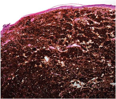
FIG. 4.14. Cutaneous malignant melanoma from a Mongolian gerbil.
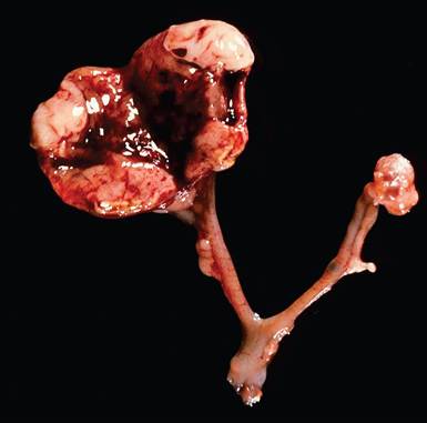
FIG. 4.16. Ovaries and uterine horns from an aged female Mongolian gerbil. The left ovary, which contains a dark red to pale granulosa cell tumor, is markedly enlarged, fleshy, and lobulated. (Source: D. Schlafer, Cornell University, Ithaca, NY. Reproduced with permission from D. Schlafer. )
that occur in different colonies of Mongolian gerbils. Cutaneous, ovarian, and adrenocortical tumors are the most commonly recognized neoplasms in this species. The most common cutaneous tumors are squamous cell carcinomas and melanomas. Melanomas (Figs. 4.14 and 4.15) tend to develop around the ear, nose, feet, and base of the tail. Squamous cell carcinomas, sebaceous adenomas, and adenocarcinomas of the ventral marking gland are relatively common in gerbils. In aged females, granulosa cell tumors appear to be the most common ovarian neoplasm. Granulosa cell tumors are frequently bilateral and vary from fleshy and lobulated to cystic masses (Fig. 4.16). Transformed granulosa cells are sometimes identifiable microscopically in the ovary in the absence of macroscopic change. On histopathology, vascular spaces surrounded by aggregations of granulosa cells
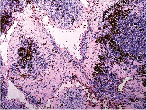
FIG. 4.15. Renal lymph node with metastasis of malignant melanoma in a Mongolian gerbil.
Also note the amyloid in interstitial areas.are the typical patterns observed (Fig. 4.17). Dysgermi- nomas, luteal cell tumors, leiomyomas, and, rarely, thecal cell carcinomas have been identified. Adrenal cortical adenomas and carcinomas also occur. Neoplasms of the lymphopoietic system are uncommon in gerbils. Leukemia with lymphoblastic infiltrates in spleen, liver, lymph nodes, and muscle was observed in an aged animal, and there are single case reports of primary cutaneous B-cell lymphoma and systemic mastocytosis. There is a relatively low incidence of tumors of the pituitary, mammary gland, and lung in the Mongolian gerbil. In a study involving other species of gerbillinae, neoplasms included squamous carcinoma of the ear, thymoma, Hodgkin-like lymphoma, uterine adenocarcinoma, adrenocortical tumors, and primary ovarian tumors. Gastric carcinomas have been produced experimentally in gerbils inoculated with H. pylori.
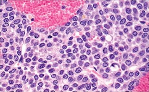
FIG. 4.17. Granulosa cell tumor in a Mongolian gerbil. Note the prominent vascular spaces.
BIBLIOGRAPHY FOR NEOPLASMS
Benitz, K.F. & Kramer, A.W. (1965) Spontaneous tumors in the Mongolian gerbil. Laboratory Animal Care 15:281-294.
Guzman-Silva, M.A. (1997) Systemic mast cell disease in a Mongolian gerbil Meriones unguiculatus: case report. Laboratory Animals 31:373-378.
Guzman-Silva, M.A. & Costa-Neves, M. (2006) Incipient spontaneous granulosa cell tumour in the gerbil, Meriones unguiculatus. Laboratory Animals 40:96-101.
Matsuoka, K. & Suzuki, J. (1995) Spontaneous tumors in the Mongolian gerbil (Meriones unguiculatus). Experimental Animals 43:755-760.
Meckley, P.E. & Zwicker, G.M. (1979) Naturally-occurring neoplasms in the Mongolian gerbil (Meriones unguiculatus). Laboratory Animals 13:203-206.
Rembert, M.S., Coleman, S.U., Klei, T.R., & Goad, M.E. (2000) Neoplastic mass in an experimental Mongolian gerbil.
Contemporary Topics in Laboratory Animal Science 39(3):34-36.Ringler, D.H., Lay, D.M., & Abrams, G.D. (1972) Spontaneous neoplasms in aging gerbillinae. Laboratory Animal Science 22:407-414.
Rowe, S.E., Simmons, J.L. Ringler, D.H., & Lay, D.M. (1974) Spontaneous neoplasms in aging Gerbillinae. Veterinary Pathology 11:38-51.
Shumaker, R.C., Paik, S.K., & Houser, W.D. (1974) Tumors in Gerbillinae: a literature review and report of a case. Laboratory Animal Science 24:688-690.
Su, Y.C., Wang, M.H., & Wu, M.F. (2001) Cutaneous B cell lymphoma in a Mongolian gerbil (Meriones unguiculatus). Contemporary Topics in Laboratory Animal Science 40(5):53- 56.
Vincent, A.L. & Ash, L.R. (1978) Further observations on spontaneous neoplasms in the Mongolian gerbil (Meriones unguicula- tus). Laboratory Animal Science 28:297-300.
Vincent, A.L., Porter, D.D., & Ash, L.R. (1975) Spontaneous lesions and parasites of the Mongolian gerbil, Meriones unguiculatus. Laboratory Animal Science 25:711-722.
Vincent, A.L., Rodrick, G.E., & Sodeman, W.A., Jr. (1979) The pathology of the Mongolian gerbil (Meriones unguiculatus): a review. Laboratory Animal Science 29:645-651.