THE CLOSTRIDIAL DISEASES
There is a group of infections in cattle all caused by one family of bacteria, the Clostridia. The clostridia are also responsible for some of the major diseases of sheep, that is pulpy kidney, lamb dysentery, enterotoxaemia, braxy etc., and they are the cause of gas gangrene in man.
In cattle there are five major syndromes, namely:tetanus
blackleg black disease botulism malignant oedema
causal agent
Clostridium tetani
Cl. chauvoei
Cl. novyi (oedematiens)
Cl. botulinum
Cl. septicum
All five diseases are similar in that infection can persist in the soil in a very resistant spore form and the bacteria grow best in the absence of air, that is, they are anaerobic.
Malignant oedema (necrotic cellulitis) is a skin disorder described in Chapter 10. Anthrax, caused by Bacillus anthracis, is a very closely related organism. It is dealt with in Chapter 11.
Tetanus
Tetanus can occur in cattle of any age and should always be considered as a possibility if an animal is showing nervous symptoms. It is generally associated with a deep and dirty wound, although in cattle the original wound may no longer be detectable by the time the symptoms of tetanus have developed. Wounds caused by an object originally coated with soil, for example penetration by a muddy nail, are especially dangerous because they take infection deep into the tissues and away from air, and anaerobic conditions such as this are exactly what the clostridia prefer. Improper application of castration rings to calves which are too old can also lead to a festering wound and tetanus. Traditionally wounds were flushed out with a solution of hydrogen peroxide. This not only kills the tetanus bacteria, but it also supplies a large quantity of oxygen to prevent their growth by destroying their anaerobic environment. Modern antiseptics have a similar effect but it is important that wounds are always cleaned first to remove dirt and soil contamination.
The use of an antibiotic aerosol after cleaning is also beneficial.Clinical signs
When infection has gained entry to the body the bacteria start to multiply and produce neurotoxins. The neurotoxins pass via the bloodstream to affect the nerve cells in the brain, and this causes either spasms or loss of function of the muscles. It is this effect which produces the clinical signs of tetanus. Initially the affected animal is dull, shows a small trembling of the muscles and is disinclined to move. A slight bloat may be noticed on the left flank because the rumen muscles have stopped working and the paralysed third eyelid passes part way across the front of the eye.
Problems with swallowing may lead to drooling and later it becomes very difficult to open the animal’s mouth, the classic lockjaw syndrome. This can be very helpful in making a specific diagnosis. As the disease progresses, stiffness becomes more apparent, then waves of muscle tremors occur, especially if the animal is excited, and its whole body may shiver uncontrollably. Eventually it is unable to stand and death follows periods of more severe muscle spasm, when all four legs and the neck become completely rigid. It is a most distressing condition to witness and as in the final stages treatment is hopeless, such animals should be humanely slaughtered.
Treatment
Your veterinary surgeon will undoubtedly be advising you on this, since treatment is extremely complex. The clostridial bacteria are easily killed by penicillin and this should prevent any further toxin from being produced, but only time and the natural defences of the animal can remove the toxins which are already present. Antiserum, containing specific antibodies to tetanus toxin, may be used and muscle relaxants and sedatives will help to overcome the muscle spasms. Animals which are not drinking should be carefully drenched (the swallowing reflex may not be functioning correctly either) and in severe cases fluids may be given intravenously.
Provided the condition is diagnosed and treated in the very early stages, it is surprising how many cattle will recover from tetanus.Prevention
There are two important aspects in the prevention of tetanus. The first is to ensure that all deep wounds are thoroughly cleaned and dressed, especially if soil contamination is a possibility.
Secondly, vaccination is highly effective and comparatively inexpensive. If animals are to be grazing areas of known tetanus risk, then they should be given two doses of vaccine at ten weeks and four weeks prior to turnout, plus an annual booster where there is a high risk. On occasions, hard swellings may develop in the skin at the site of vaccination. These will slowly disappear without treatment. Sometimes large numbers of animals from a single group develop tetanus over a short period of time and no cause is found. This is known as idiopathic tetanus. If a single animal is affected it is therefore a wise precaution to immediately vaccinate the remainder of the group to prevent further cases.
Blackleg
Blackleg most commonly affects cattle approximately six to eighteen months old and it is almost always a disease of grazing animals or of housed animals which have previously grazed infected pastures. It is caused by the bacterium Clostridium chauvoei, which is present in the soil and is eaten during grazing. The factors which lead to the development of blackleg in an animal carrying spores in its muscles are unknown. It is possible to dose calves with Cl. chauvoei spores and produce no effect. Muscle bruising, e.g. trauma or ‘bulling’ activity, may be important, as bruising can produce anaerobic conditions in muscles which just happen to be carrying blackleg spores. However, soil or pasture must be implicated because disease seems to be more prevalent on certain fields and especially fields which flood.
Clinical signs
It is unlikely that you will see anything but a dead animal, because the disease is so acute.
However, on occasions you may witness an animal which is very dull, standing apart from the others and perhaps panting. Characteristically there will be a swelling somewhere in the muscles where the bacteria are growing, and this is seen in both the live and the dead animal as an enlargement under the skin, often along the back or in the hind legs. If squeezed, a cracking sound is heard, due to the massive accumulation of gas produced by the bacteria. After death the affected muscles have a butyric or rancid smell and are much darker in colour - hence the name blackleg.Plate 4.26 shows a typical example. Note the very dark muscle in the right leg compared with normal muscle in the left. This animal was in a field bordered by the River Severn. For many years the farmer had vaccinated his cattle prior to turnout, but that year he simply forgot. The animal was seen alive, but acutely lame with a huge swelling in the muscles of the right hind leg. Although massive doses of penicillin were given, it died within a few hours. In some animals only the heart is affected. This could be missed at post-mortem.
Treatment and control
Treatment is rarely possible, although if a live affected animal is seen, massive doses of penicillin may be effective. Vaccination is the only means of prevention and a combined blackleg and tetanus vaccine is commonly used.
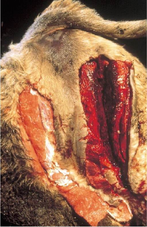
Plate 4.26. Blackleg, a clostridial infection of the muscles. The right hind is swollen and the muscle is very dark compared with the normal left leg.
Black Disease (Infectious Necrotic Hepatitis)
This is certainly not a common disorder, but may occasionally be seen in grazing calves. The organism Cl. novyi (oedematiens) is ingested with soil-contaminated food and multiplies in the liver, where it causes a type of ‘gas gangrene’ similar to blackleg, and, again, very rapid death. If a live affected animal is seen, then penicillin would be the drug of choice for treatment.
Control is by vaccination and this is highly effective. Liver fluke larvae migrating across the liver are thought to cause damage which encourages the growth of Cl. novyi, so fluke control (Chapter 13) is also important in prevention. Combined tetanus, blackleg and black disease vaccines are available and cost little more than the tetanus vaccine alone. Vaccination would be advisable in fluke areas.
Botulism
This is a rare disease of cattle in the British Isles, although it occurs more commonly overseas. Botulism is an intoxication, not an infection. The bacteria, Cl. botulinum, may be present in the gut of normal healthy animals and cause no problems. After death, however, the bacteria may multiply rapidly and produce a toxin. If other cattle then consume (probably inadvertently via contaminated feed or water) part of the dead carcase containing the toxin, they will develop a progressive paralysis, eventually causing death from loss of function of the respiratory muscles. The toxin of Cl. botulinum is one of the most deadly substances known to man, with minute quantities being fatal. Outbreaks in the UK are usually associated with cattle grazing pasture which has been fertilised with chicken manure containing dead chickens.
Ryegrass Staggers
Ryegrass staggers is not a clostridial infection, but it is included here because it is seen in grazing animals and it produces nervous signs and peculiarities of gait which could be confused with the early stages of tetanus or botulism. Affected animals are normal when resting, but when moved they may tremble slightly, drag one or both hind legs behind them, or collapse if their front legs give way. Sheep are more commonly affected than cattle. The condition is seen after a very dry summer, when cattle or sheep are grazing perennial ryegrass pastures tight to the ground. It is caused by ingestion of the toxin Lolitrem B, produced by the fungus Accremonium lolii. If cattle are removed from the perennial ryegrass pasture they recover within a few days without treatment.
More on the topic THE CLOSTRIDIAL DISEASES:
- OTHER CLOSTRIDIAL DISEASES IN WILDLIFE
- Clostridial Diseases
- The various cardiovascular diseases observed in HIV-infected patients and widely described in the literature have been predominantly coronary and peripheral arterial diseases (PAD) and remain poorly known.
- Clostridium difficile and Clostridium perfringens: Clostridial Enteropathy
- Clostridium difficile, Clostridium perfringens, and Clostridium spiroforme: Clostridial Enteropathy
- BIBLIOGRAPHY FOR NONINFECTIOUS DISEASES
- INTERSTITIAL LUNG DISEASES
- WHY STUDY WILDLIFE DISEASES
- VECTOR-BORNE VIRAL DISEASES
- BIBLIOGRAPHY FOR PARASITIC DISEASES
- Bibliography for parasitic diseases
- BIBLIOGRAPHY FOR PARASITIC DISEASES
- GENERAL REFERENCES ON DISEASES OF MICE
- 6 DISEASES BY CLINICAL PRESENTATIONS, MAMMALS
- ROLE OF DISEASES IN WILDLIFE POPULATIONS
- 7 DISEASES BY CLINICAL PRESENTATIONS, BIRDS
- RESPIRATORY DISEASES
- Notifiable Diseases